- The Neuron

Содержание
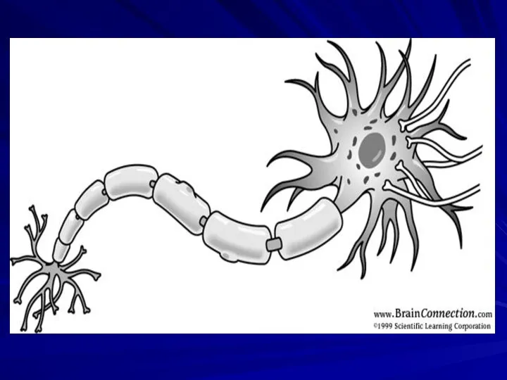
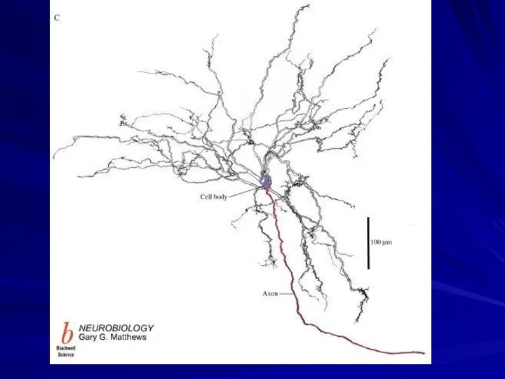
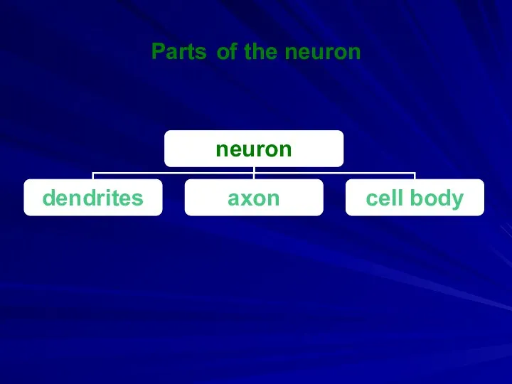
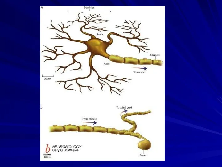
- 5. Parts of the neuron
- 6. Dendrites The dendrites are the part of the neuron that receive signals from other neurons. The
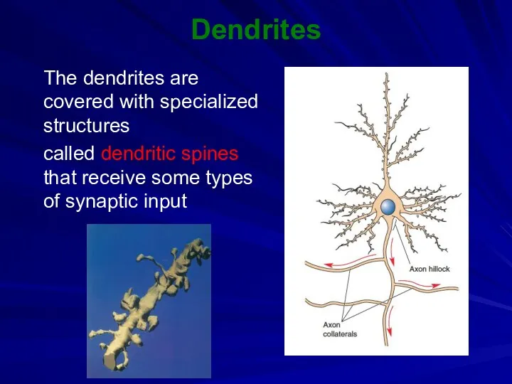
- 7. Dendrites The dendrites are covered with specialized structures called dendritic spines that receive some types of
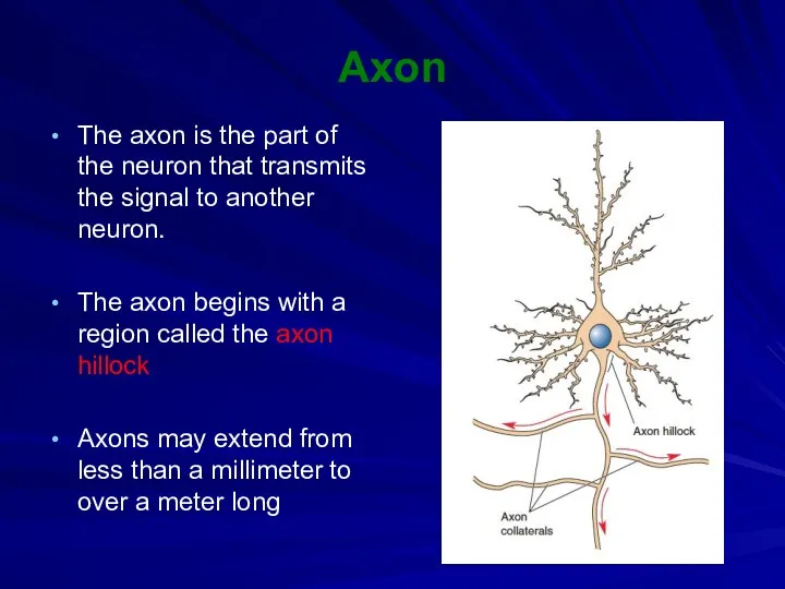
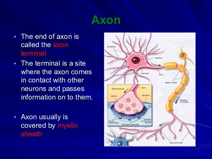
- 9. Axon The axon is the part of the neuron that transmits the signal to another neuron.
- 10. Axon The end of axon is called the axon terminal The terminal is a site where
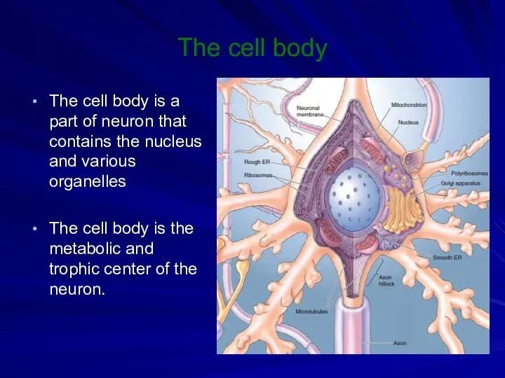
- 11. The cell body The cell body is a part of neuron that contains the nucleus and
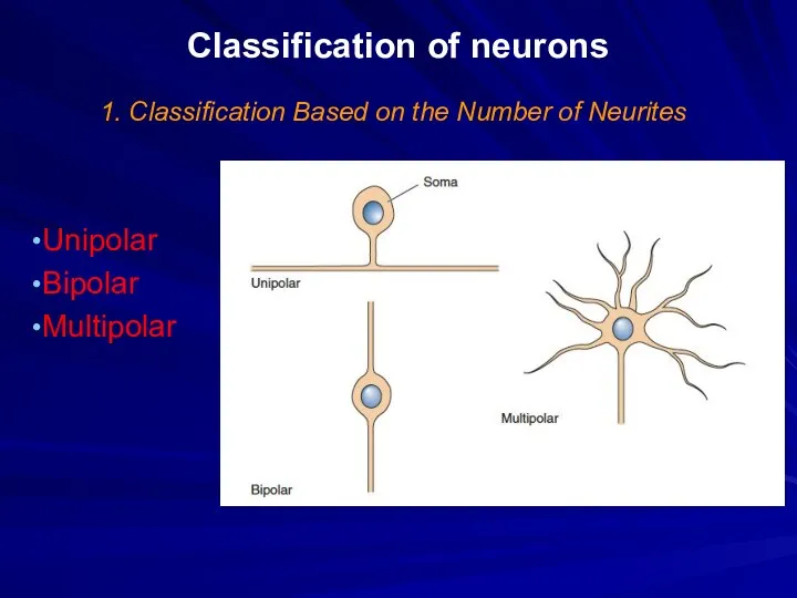
- 13. Classification of neurons 1. Classification Based on the Number of Neurites Unipolar Bipolar Multipolar
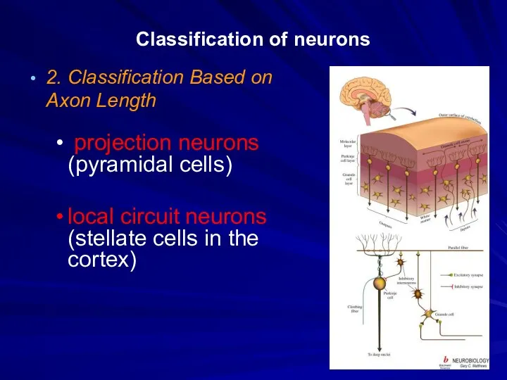
- 14. Classification of neurons 2. Classification Based on Axon Length projection neurons (pyramidal cells) local circuit neurons
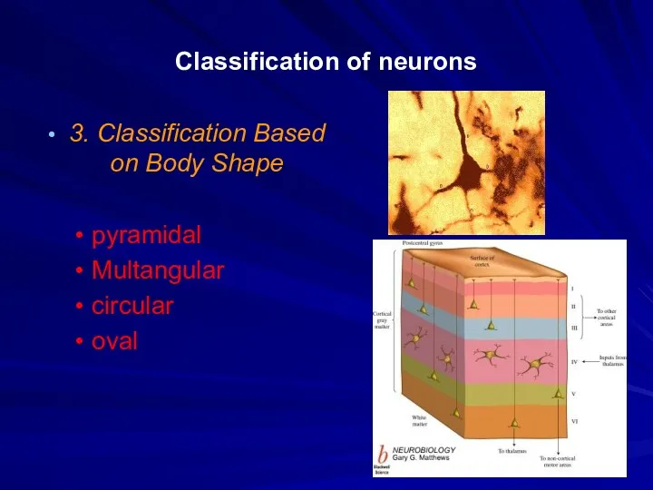
- 16. Classification of neurons 3. Classification Based on Body Shape pyramidal Multangular circular oval
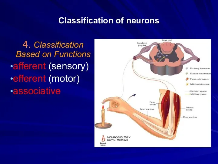
- 17. Classification of neurons 4. Classification Based on Functions afferent (sensory) efferent (motor) associative
- 18. Synapses Synapse the specialized junction where one part of a neuron contacts and communicates with another
- 20. Classification of Synapses 1. The place of junction neuromuscular junction neuro-neural junction neuro-glandular junction
- 21. Classification of Synapses 2. Synaptic arrangements in the CNS axodendritic synapse axosomatic synapse axoaxonic synapse
- 23. Classification of Synapses 3. Based of type of influence excitatory inhibitory
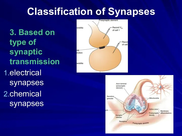
- 24. Classification of Synapses 3. Based on type of synaptic transmission electrical synapses chemical synapses
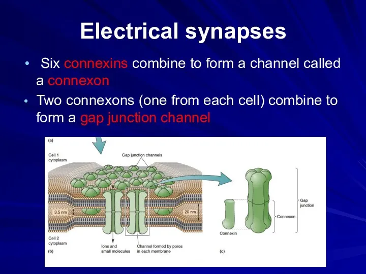
- 25. Electrical synapses Six connexins combine to form a channel called a connexon Two connexons (one from
- 26. Chemical synapses Axon terminal Presynaptic membrane Postsynaptic membrane Synaptic cleft Synaptic vesicles Postsynaptic receptors
- 28. Nerve fibers
- 29. Classification of nerve fibers unmyelinated fibers myelinated fibers
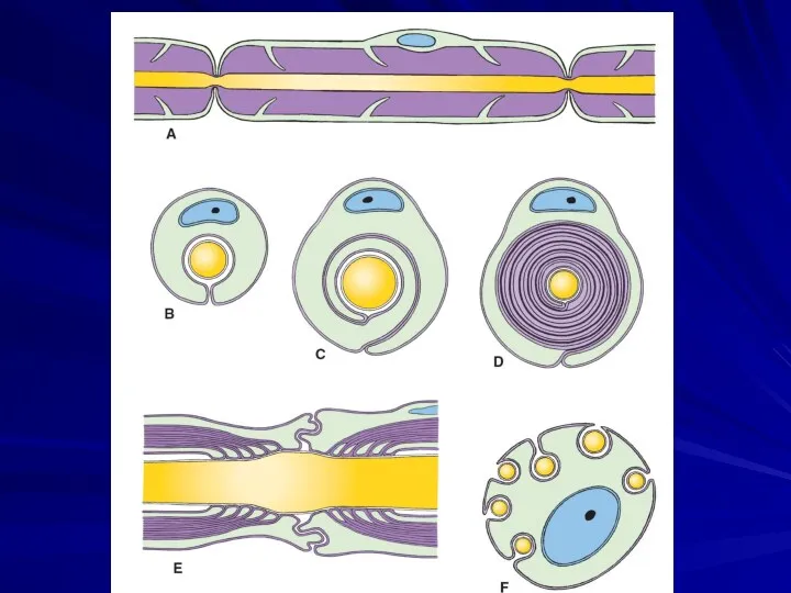
- 30. Myelin sheath Myelin sheath is a fatty white substance that surrounds the axon of nerve fibers,
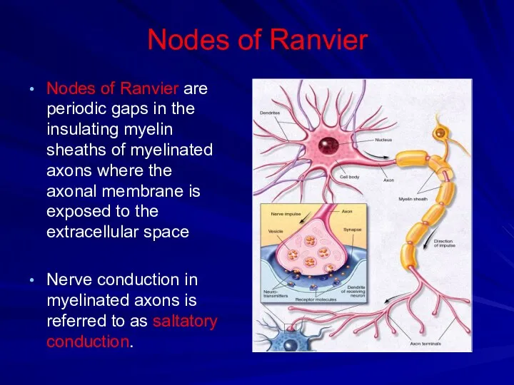
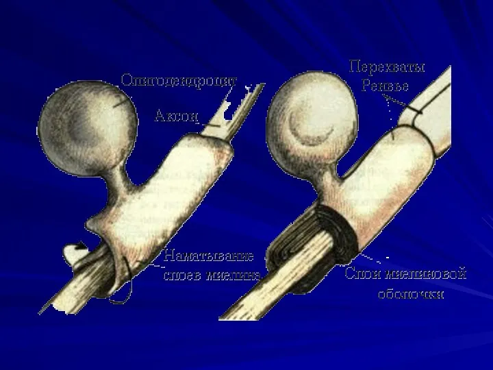
- 32. Nodes of Ranvier Nodes of Ranvier are periodic gaps in the insulating myelin sheaths of myelinated
- 35. Glial cells Glial cells (neuroglia or glia) are non-neuronal cells that maintain homeostasis, form myelin, and
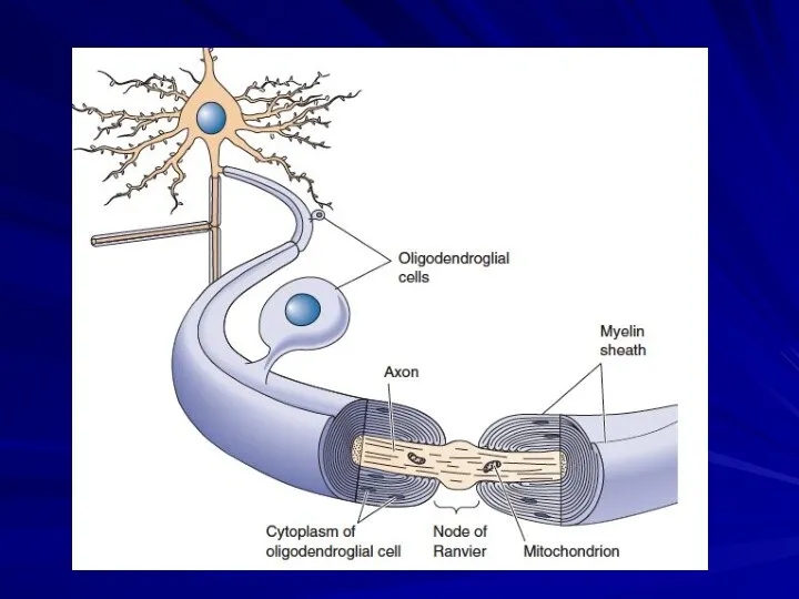
- 36. Classification of glial cells Schwann cells supply the myelin for the peripheral nervous system Oligodendrocytes myelinate
- 38. Скачать презентацию
Parts of the neuron
Parts of the neuron
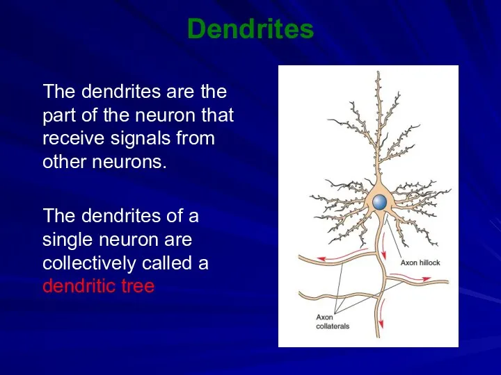
Dendrites
The dendrites are the part of the neuron that receive
Dendrites
The dendrites are the part of the neuron that receive
Dendrites
The dendrites are covered with specialized structures
called dendritic spines that
Dendrites
The dendrites are covered with specialized structures
called dendritic spines that
Axon
The axon is the part of the neuron that transmits
Axon
The axon is the part of the neuron that transmits
Axon
The end of axon is called the axon terminal
The terminal
Axon
The end of axon is called the axon terminal
The terminal
The cell body
The cell body is a part of neuron
The cell body
The cell body is a part of neuron
Classification of neurons
1. Classification Based on the Number of Neurites
Unipolar
Bipolar
Multipolar
Classification of neurons
1. Classification Based on the Number of Neurites
Unipolar
Bipolar
Multipolar
Classification of neurons
2. Classification Based on Axon Length
projection neurons (pyramidal
Classification of neurons
2. Classification Based on Axon Length
projection neurons (pyramidal
Classification of neurons
3. Classification Based on Body Shape
pyramidal
Multangular
circular
oval
Classification of neurons
3. Classification Based on Body Shape
pyramidal
Multangular
circular
oval
Classification of neurons
4. Classification Based on Functions
afferent (sensory)
efferent (motor)
associative
Classification of neurons
4. Classification Based on Functions
afferent (sensory)
efferent (motor)
associative
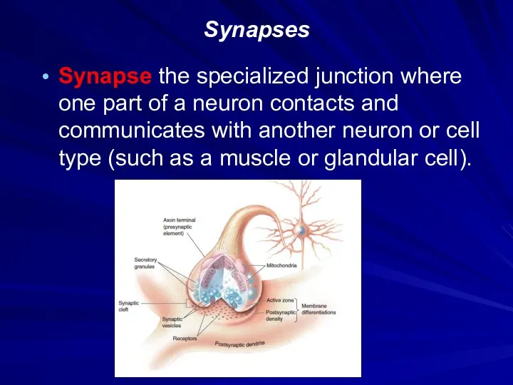
Synapses
Synapse the specialized junction where one part of a neuron contacts
Synapses
Synapse the specialized junction where one part of a neuron contacts

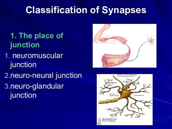
Classification of Synapses
1. The place of junction
neuromuscular junction
neuro-neural junction
neuro-glandular junction
Classification of Synapses
1. The place of junction
neuromuscular junction
neuro-neural junction
neuro-glandular junction
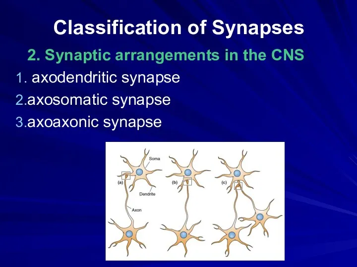
Classification of Synapses
2. Synaptic arrangements in the CNS
axodendritic synapse
axosomatic synapse
axoaxonic
Classification of Synapses
2. Synaptic arrangements in the CNS
axodendritic synapse
axosomatic synapse
axoaxonic

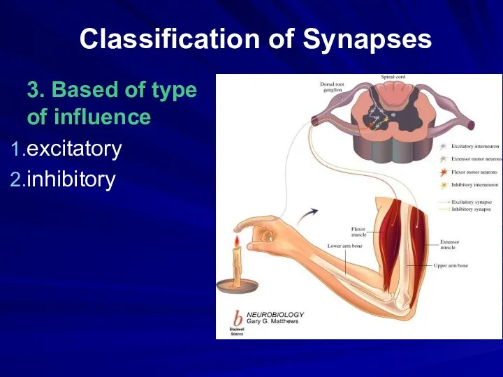
Classification of Synapses
3. Based of type of influence
excitatory
inhibitory
Classification of Synapses
3. Based of type of influence
excitatory
inhibitory
Classification of Synapses
3. Based on type of synaptic transmission
electrical synapses
chemical synapses
Classification of Synapses
3. Based on type of synaptic transmission
electrical synapses
chemical synapses
Electrical synapses
Six connexins combine to form a channel called a
Electrical synapses
Six connexins combine to form a channel called a
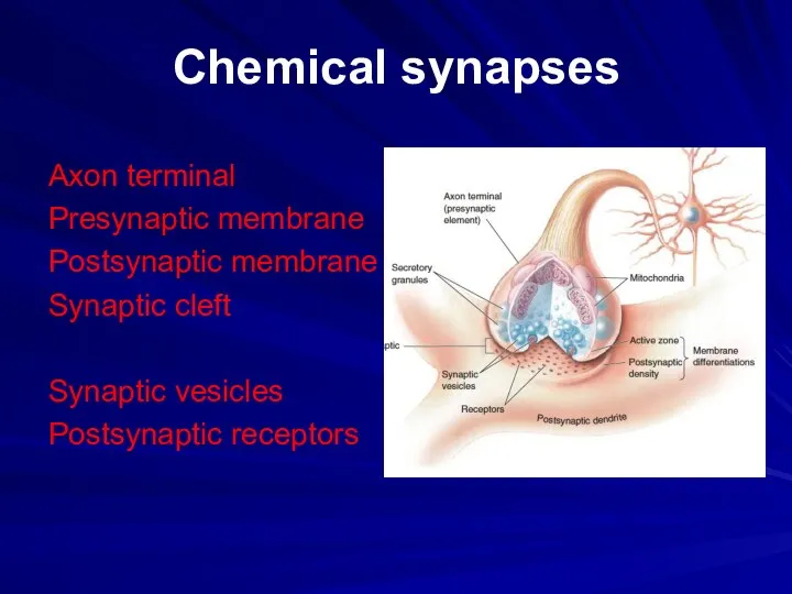
Chemical synapses
Axon terminal
Presynaptic membrane
Postsynaptic membrane
Synaptic cleft
Synaptic vesicles
Postsynaptic receptors
Chemical synapses
Axon terminal
Presynaptic membrane
Postsynaptic membrane
Synaptic cleft
Synaptic vesicles
Postsynaptic receptors
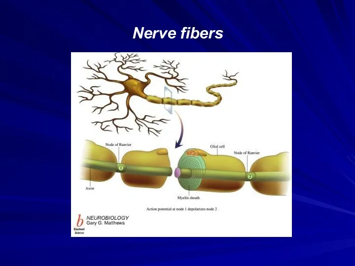
Nerve fibers
Nerve fibers
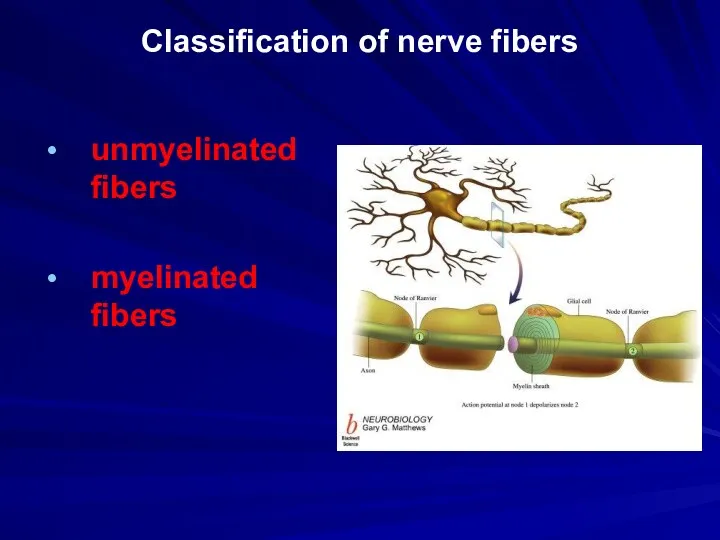
Classification of nerve fibers
unmyelinated fibers
myelinated fibers
Classification of nerve fibers
unmyelinated fibers
myelinated fibers
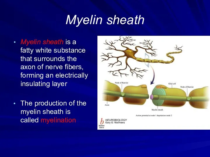
Myelin sheath
Myelin sheath is a fatty white substance that surrounds the
Myelin sheath
Myelin sheath is a fatty white substance that surrounds the
Nodes of Ranvier
Nodes of Ranvier are periodic gaps in the insulating
Nodes of Ranvier
Nodes of Ranvier are periodic gaps in the insulating
Glial cells
Glial cells (neuroglia or glia) are non-neuronal cells that maintain
Glial cells
Glial cells (neuroglia or glia) are non-neuronal cells that maintain
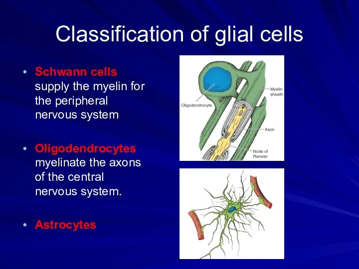
Classification of glial cells
Schwann cells supply the myelin for the peripheral
Classification of glial cells
Schwann cells supply the myelin for the peripheral
 Способы словообразования. Частотные отрезки, наиболее часто употребляемые в названиях медицинских терминов
 Общественное здоровье. (Лекция 1.1)
 Конъюнктивиты
 ВБИ
ВБИ Денсаулық сақтау ұйымында сапа менеджмент жүйесі
 Тромботический синдром
 Теория рационального питания. Гигиенические требования к рациональному питанию человека
 Нейропатическая боль. Определение
 Моральні та правові аспекти лікарської таємниці
 Тамақтанудың ауыр бұзылысы
 Өзгергіштік
 Мужское бесплодие
 Патофизиология внешнего дыхания
 Принципы клинической эпидемиологии
 Роль труда, спорта, социальных и биологических факторов на строение костей
 Синдром слабости синусового узла
 Радиоактивті индикаторларды халық шаруашылығының әртүрлі салаларында қолданылуы
 Формы клинического течения инсульта
 Blood vessels pathology. (Subject 14)
 Бронхиты. Бронхиальная астма. Эмфизема
 Нарушения пигментации кожи (дисхромии)
 Использование эндоскопических методов исследования в акушерстве и гинекологии
 Болезни дыхательной системы и их предупреждение
 Острый панкреатит и его лечение
 Участие медицинской сестры в инструментальных методах исследования
 Бір бөлімді және көп бөлімді қарындардың қимылы.Асқорыту ерекшеліктерінің реттелуі
 Основы токсикологии. (Лекция 1)
 Окислительный стресс