- Traumatic injuries of kidneys, ureter, bladder

Содержание
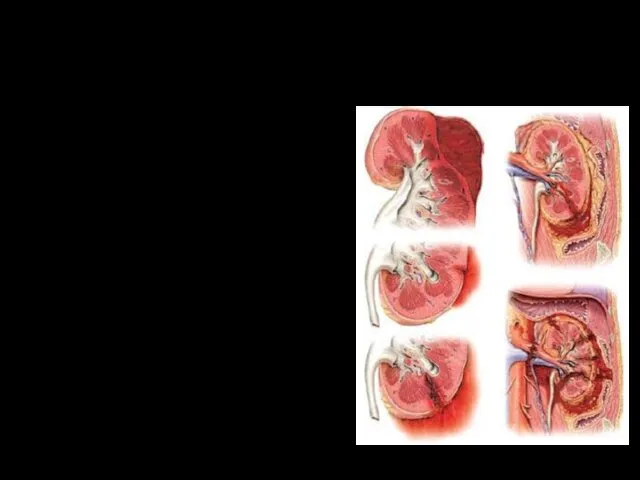
- 2. Closed kidney damage - Damage to the fat and fibrous capsules with the formation of a
- 3. Mechanism of closed kidney damage Causes: Blunt blunt objects Shaking Pressure The degree of damage depends
- 4. Open kidney damage By the type of the hurting projectile: firearms (bullet, shrapnel, explosive); non-fireable In
- 5. Iatrogenic exposure Retrograde pyelography Puncture Shockwave remote lithotripsy
- 6. Clinical manifestations Dysuria Symptoms of peritoneal irritation Nausea Vomiting Fever Gastrointestinal dysfunction Lumbar pain Hematuria Swelling
- 7. Three degrees of severity Mild kidney injury - the general condition of the victim is poorly
- 8. Diagnostics On examination: Hematoma, swelling in the lumbar region Local muscle tension Rib fractures Paleness of
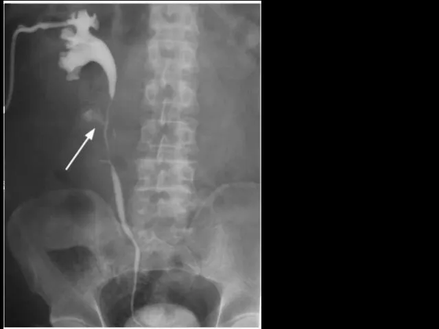
- 9. Contrast radiography
- 10. Rupture of the left kidney
- 11. Treatment Stopping bleeding Bed rest 10-15 days Control of hemodynamics and hematocrit Preventive parenteral administration of
- 12. Damage of the ureters Ureters are rarely damaged due to elasticity, displaceability and location. Iatrogenic damage
- 13. Classification By type: Closed ureteral injury (subcutaneous). Open ureteral injury (wound). By the nature: An isolated
- 14. Diagnostics Diagnosis is based on an analysis of the circumstances and mechanism of injury, clinical manifestations
- 15. Antegrade pyeloutraprogram
- 16. Differential diagnostics To distinguish between injuries of the ureter and bladder, use the method of filling
- 17. Bladder damage Causes: blunt or penetrating injury leading to rupture Mechanism Blunt blow to full bladder;
- 18. Closed (with integer integument): injury; incomplete rupture (external and internal); complete break; two-stage rupture of the
- 19. Clinical manifestations Intraperitoneal Pain over pubis Anuria Signs of peritonitis Bloating Symptom "Vanka-Vstanki" Extraperitoneal Pain over
- 20. Diagnostics Catheterization Zeldovich positive symptom (inconsistency between the injected and exiting fluid from the catheter) AS
- 21. Retrograde cystogram. Extraperitoneal bladder rupture
- 22. Intraperitoneal bladder rupture
- 23. Flow of contrast fluid into paravesical space
- 24. Treatment Conservative Surgical Bed rest Uroseptics and antibiotics Hemostatic therapy NSAIDs Cold compresses on the stomach
- 25. Drainage by Buyalsky-McWorthier
- 27. Скачать презентацию
Closed kidney damage
- Damage to the fat and fibrous capsules with
Closed kidney damage
- Damage to the fat and fibrous capsules with

Mechanism of closed kidney damage
Causes:
Blunt blunt objects
Shaking
Pressure
The degree of
Mechanism of closed kidney damage
Causes:
Blunt blunt objects
Shaking
Pressure
The degree of
Open kidney damage
By the type of the hurting projectile:
firearms (bullet, shrapnel,
Open kidney damage
By the type of the hurting projectile:
firearms (bullet, shrapnel,
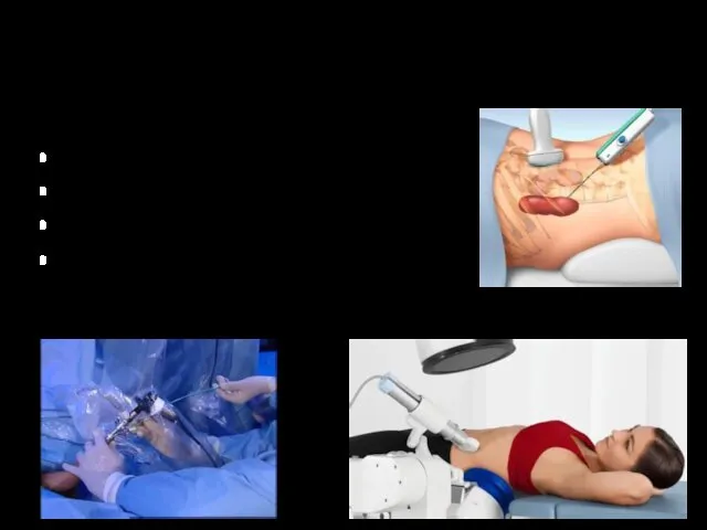
Iatrogenic exposure
Retrograde pyelography
Puncture
Shockwave remote lithotripsy
Iatrogenic exposure
Retrograde pyelography
Puncture
Shockwave remote lithotripsy

Clinical manifestations
Dysuria
Symptoms of peritoneal irritation
Nausea
Vomiting
Fever
Gastrointestinal dysfunction
Lumbar pain
Hematuria
Swelling
Clinical manifestations
Dysuria
Symptoms of peritoneal irritation
Nausea
Vomiting
Fever
Gastrointestinal dysfunction
Lumbar pain
Hematuria
Swelling

Three degrees of severity
Mild kidney injury - the general condition of
Three degrees of severity
Mild kidney injury - the general condition of

Diagnostics
On examination:
Hematoma, swelling in the lumbar region
Local muscle tension
Rib fractures
Paleness of
Diagnostics
On examination:
Hematoma, swelling in the lumbar region
Local muscle tension
Rib fractures
Paleness of
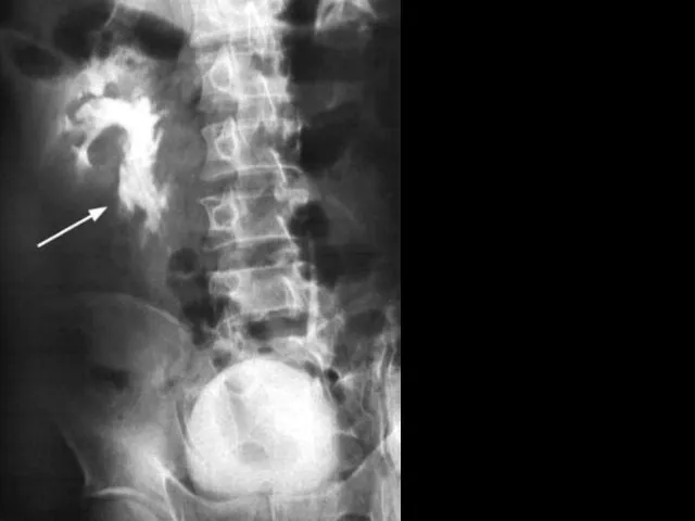
Contrast radiography
Contrast radiography
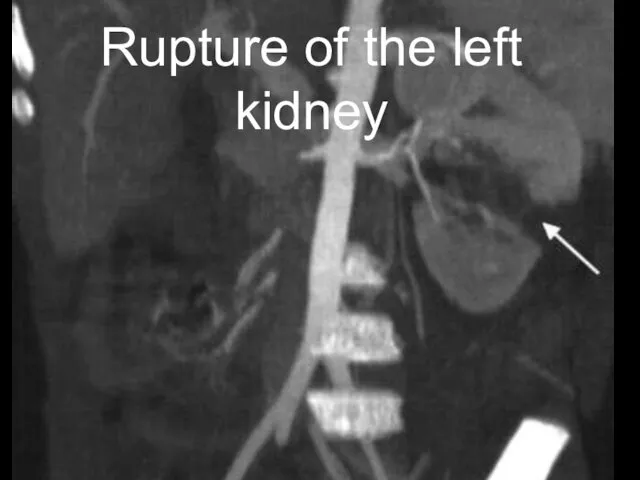
Rupture of the left kidney
Rupture of the left kidney

Treatment
Stopping bleeding
Bed rest 10-15 days
Control of hemodynamics and hematocrit
Preventive parenteral administration
Treatment
Stopping bleeding
Bed rest 10-15 days
Control of hemodynamics and hematocrit
Preventive parenteral administration

Damage of the ureters
Ureters are rarely damaged due to elasticity, displaceability
Damage of the ureters
Ureters are rarely damaged due to elasticity, displaceability

Classification
By type:
Closed ureteral injury (subcutaneous).
Open ureteral injury (wound).
By the nature:
An isolated
Classification
By type:
Closed ureteral injury (subcutaneous).
Open ureteral injury (wound).
By the nature:
An isolated

Diagnostics
Diagnosis is based on an analysis of the circumstances and mechanism
Diagnostics
Diagnosis is based on an analysis of the circumstances and mechanism
Antegrade pyeloutraprogram
Antegrade pyeloutraprogram

Differential diagnostics
To distinguish between injuries of the ureter and bladder, use
Differential diagnostics
To distinguish between injuries of the ureter and bladder, use

Bladder damage
Causes: blunt or penetrating injury leading to rupture
Mechanism
Blunt blow to
Bladder damage
Causes: blunt or penetrating injury leading to rupture
Mechanism
Blunt blow to

Closed (with integer integument):
injury;
incomplete rupture (external and internal);
complete break;
two-stage rupture of
Closed (with integer integument):
injury;
incomplete rupture (external and internal);
complete break;
two-stage rupture of
Clinical manifestations
Intraperitoneal
Pain over pubis
Anuria
Signs of peritonitis
Bloating
Symptom "Vanka-Vstanki"
Extraperitoneal
Pain over the bosom and
Clinical manifestations
Intraperitoneal
Pain over pubis
Anuria
Signs of peritonitis
Bloating
Symptom "Vanka-Vstanki"
Extraperitoneal
Pain over the bosom and

Diagnostics
Catheterization
Zeldovich positive symptom (inconsistency between the injected and exiting fluid from
Diagnostics
Catheterization
Zeldovich positive symptom (inconsistency between the injected and exiting fluid from
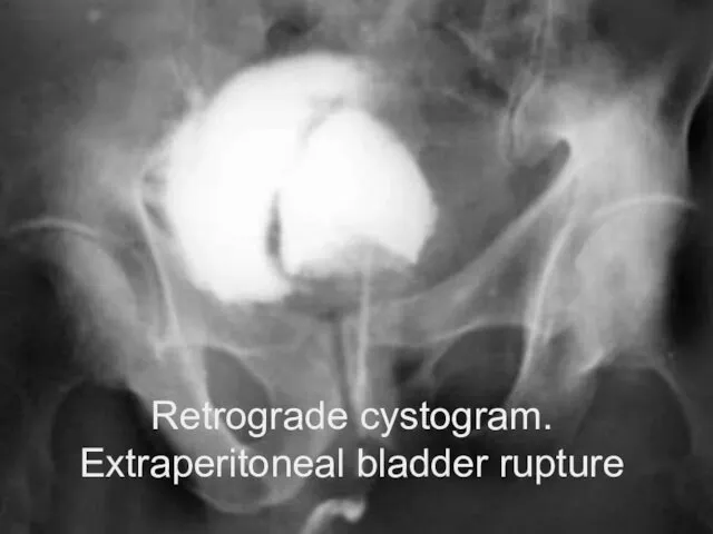
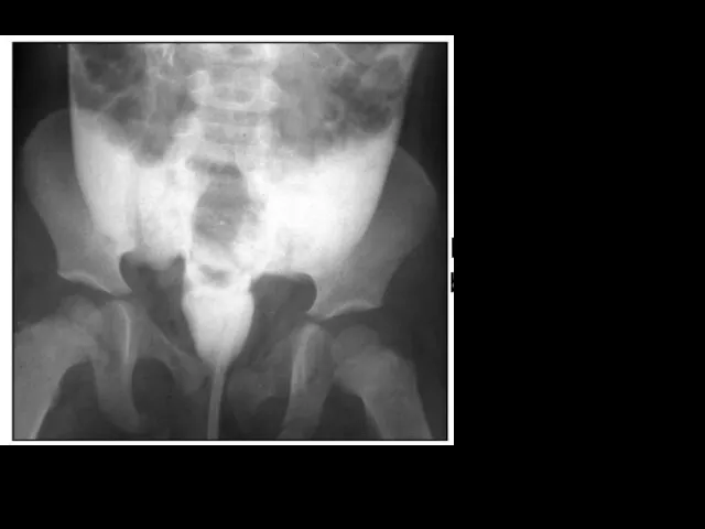
Retrograde cystogram. Extraperitoneal bladder rupture
Retrograde cystogram. Extraperitoneal bladder rupture
Intraperitoneal bladder rupture
Intraperitoneal bladder rupture
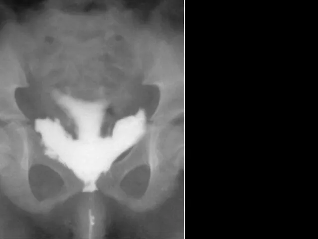
Flow of contrast fluid into paravesical space
Flow of contrast fluid into paravesical space

Treatment
Conservative Surgical
Bed rest
Uroseptics and antibiotics
Hemostatic therapy
NSAIDs
Cold compresses on the stomach
Catheterization
Restoring
Treatment
Conservative Surgical
Bed rest
Uroseptics and antibiotics
Hemostatic therapy
NSAIDs
Cold compresses on the stomach
Catheterization
Restoring
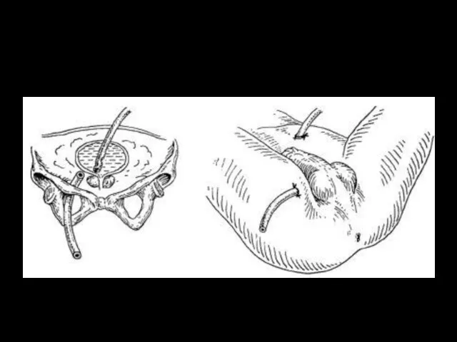
Drainage by Buyalsky-McWorthier
Drainage by Buyalsky-McWorthier
 Микотоксины. Действие микотоксинов в истории
 Общие принципы лечения абсцессов и флегмон лица и шеи. Физиотерапия и реабилитация больных
 Диспансерное наблюдение за детьми с хроническими заболеваниями
 Хронические расстройства питания у детей
 Наследственные заболевания человека
 Геморрагический шок
 Гипогликемическая и гипергликемическая комы
 Пути введения лекарственных средств
 Обзор и принципы реанимации новорожденных
 Современные алгоритмы лечения сахарного диабета 2 типа
 Нейропсихологическая диагностика
 Общая характеристика группы инфекционных болезней с воздушнокапельным механизмом передачи. Грипп
 Вагинальные инфекции при беременности
 Federal State Educational Institution of Higher Education
 Синдром наличия жидкости и газа в плевральной полости. Плевриты
 Холера. Эпидемиология
 Хронический пылевой бронхит
 Легочное сердце
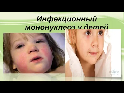 Инфекционный мононуклеоз у детей
 Понятие гиподинамии, гипердинамии
 Действия ассистента, осуществляемые до прихода врача-стоматолога, после прихода врача-стоматолога и после окончания лечения
 Митральные пороки сердца
 Критерии и качества стоматологических материалов. Система международных и национальных стандартов
 Введение в венерологию. История развития венерологии. Инфекции, передающиеся половым путем
 Доказательная профилактика. Скрининговые программы
 Endocrine system
 Современные подходы к лечению эндометриоидных кист яичников
 Дисфункционалдық жатырдан қан кету