- Tumor pathogenesis. (Lecture 7)

Содержание
- 2. Growth factors Epidermal growth factor Endothelial growth factor Fibroblast growth factor Platelet-derived growth factor Nerve growth
- 3. Genes controlling growth The genes which are controlling growth factors synthesis are named proto-oncogenes. If gene
- 4. Mutagenic factors chemical - pro-carcinogens (substances that can convert to carcinogens) and carcinogens. Aflatoxin (from fungus
- 5. Mutagenic factors physical (ionizing radiation, ultraviolet rays); biological - viruses. The viral particles can carry a
- 6. Epigenetic carcinogenesis Transformation of the normal cel to tumor one without mutations by stimulating mitosis may
- 7. The main causes of cancer Smoking, Dietary imbalances (excess fat and calories; inadequate intake of fruits,
- 8. Role of host factors and environment Microsomal enzymes in the liver degrade a large part of
- 9. Definitions Neoplasia A pathologic process in which a permanent alteration in a cell’s growth controlling mechanism
- 10. General pathogenesis of tumor mutation CLONE mutation
- 11. General Pathogenesis of tumors Stage 1. INITIATION. Normal cell under the effect of etiological factors obtains
- 12. General Pathogenesis of tumors Stage 3. TUMOR PROGRESSION. Regulatory systems of the organism affect the multiplying
- 13. General pathogenesis of tumors Initiation Promotion Progression
- 14. Types of neoplasms Benign – less autonomy, usually not invasive, does not metastasize, and generally produces
- 15. Common characteristics of neoplasms Neoplasia is an irreversible new growth Growth controlling mechanisms are impaired in
- 16. Common characteristics of neoplasms Absence of cell division limit. Normal cells have division limit Tumor cells
- 17. Common characteristics of neoplasms Inadequate Differentiation Neoplastic cells do not become as specialized as do normal
- 18. Characteristics Unique to Malignant Neoplasms Loss of differentiation is known as “anaplasia.” Anaplasia is seen only
- 19. Characteristics Unique to Malignant Neoplasms Morphological anaplasia. Pleomorphism (pleo- = many; -morphism = shape). Abnormally large
- 20. Characteristics Unique to Malignant Neoplasms Biochemical anaplasia. Carbohydrate atypia –shift to anaerobic glycolysis. negative Paster’s effect
- 21. Characteristics Unique to Malignant Neoplasms Functional anaplasia Normal cells stop their division when they contact surrounding
- 22. Characteristics Unique to Malignant Neoplasms Loss of polaruty and specialized functions Normal differentiated cells are polar
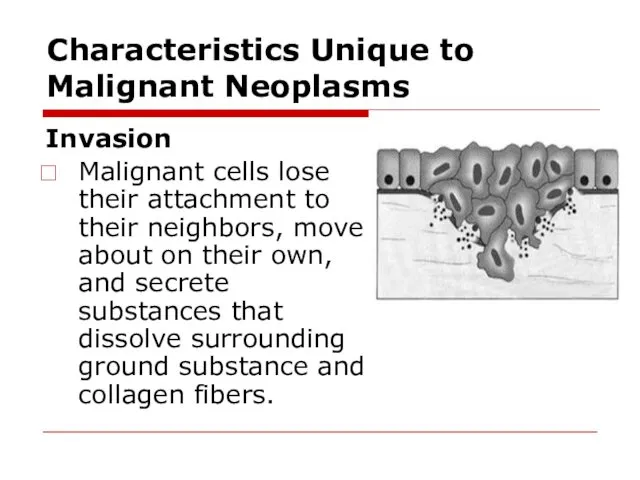
- 23. Characteristics Unique to Malignant Neoplasms Invasion Malignant cells lose their attachment to their neighbors, move about
- 24. Characteristics Unique to Malignant Neoplasms Invasive malignancies are difficult to eradicate. There is no line of
- 25. Characteristics Unique to Malignant Neoplasms Metastasis The tendency of malignant neoplasms to spread far is called
- 26. Invasion and metastasis of malignant tumor cells Tumor cell invasion. The sequence of events: detachment of
- 27. Invasion and metastasis of malignant tumor cells Tumor Cell Embolization. Malignant tumor cells may invade lymphatic
- 28. Invasion and metastasis of malignant tumor cells Tumor Cell Extravasation. The sequence of mechanisms includes: adhesion
- 29. Metastasis of tumor cells Malignant tumor cells may spread by three major routes: lymphatics, blood vessels,
- 30. Other Differences between Benign and Malignant Neoplasms Benign Neoplasms growth pattern is known as expansive growth.
- 31. Other Differences between Benign and Malignant Neoplasms Malignant neoplasms grow rapidly and have the capacity to
- 32. Summary of differences
- 33. Summary of differences
- 34. How tumor cell escape immune surveilance low immunogenicity of tumor antigens constant modification of tumor antigens
- 35. Organism defense against tumor anticarcinogenic mechanisms antimutational mechanisms anticellular mechanisms
- 36. Organism defense against tumor Anticarcinogenic mechanisms - braking of carcinogens entrance, their inactivation and elimination from
- 37. Organism defense against tumor Antimutational mechanisms provide revealing, elimination or inhibition of oncogenes activity with the
- 38. Organism defense against tumor TNF is released primarily by WBC and endothelium. Stimulating of the corticotropin
- 39. Interrelations between the host organism and the tumor Peculiarities of tumor Location is of critical importance
- 40. Interrelations between the host organism and the tumor Cancer Cachexia is a progressive loss of body
- 41. Interrelations between the host organism and the tumor The paraneoplastic syndromes: hypercalcemia, Cushing's syndrome, and nonbacterial
- 42. Cancer grading and staging The cancer may be classified as grade: I, II, III, or IV,
- 43. Cancer grading and staging Staging of a malignant neoplasm assesses its amount of invasion and metastasis.
- 44. Treatment of Neoplasms Surgery.” Situations in which little surrounding tissue needs to be sacrificed is called
- 46. Скачать презентацию
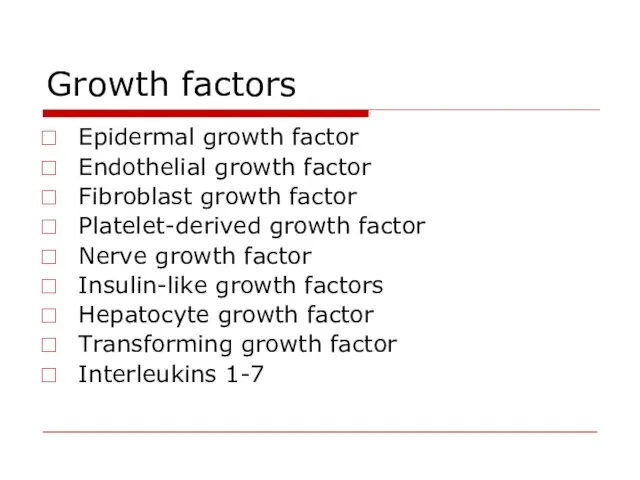
Growth factors
Epidermal growth factor
Endothelial growth factor
Fibroblast growth factor
Platelet-derived growth
Growth factors
Epidermal growth factor
Endothelial growth factor
Fibroblast growth factor
Platelet-derived growth
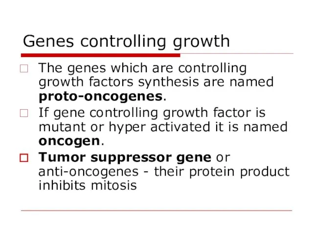
Genes controlling growth
The genes which are controlling growth factors synthesis
Genes controlling growth
The genes which are controlling growth factors synthesis
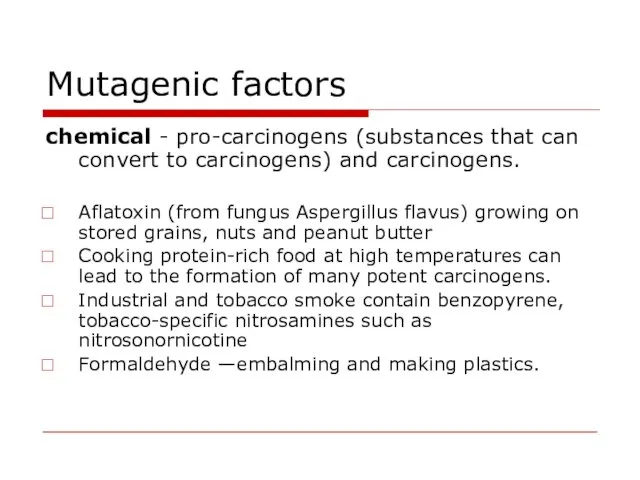
Mutagenic factors
chemical - pro-carcinogens (substances that can convert to carcinogens) and
Mutagenic factors
chemical - pro-carcinogens (substances that can convert to carcinogens) and
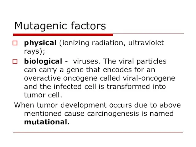
Mutagenic factors
physical (ionizing radiation, ultraviolet rays);
biological - viruses. The viral
Mutagenic factors
physical (ionizing radiation, ultraviolet rays);
biological - viruses. The viral
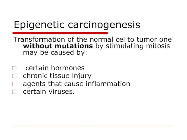
Epigenetic carcinogenesis
Transformation of the normal cel to tumor one without
Epigenetic carcinogenesis
Transformation of the normal cel to tumor one without
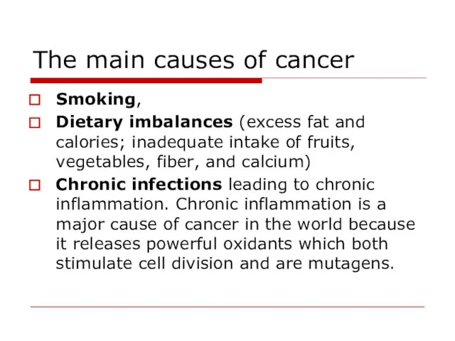
The main causes of cancer
Smoking,
Dietary imbalances (excess fat and calories;
The main causes of cancer
Smoking,
Dietary imbalances (excess fat and calories;
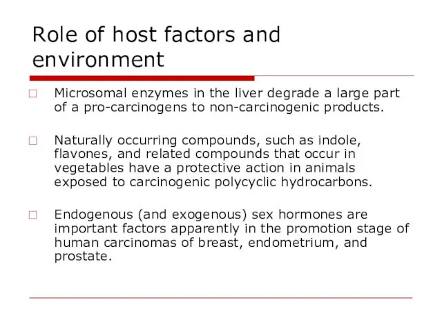
Role of host factors and environment
Microsomal enzymes in the liver degrade
Role of host factors and environment
Microsomal enzymes in the liver degrade
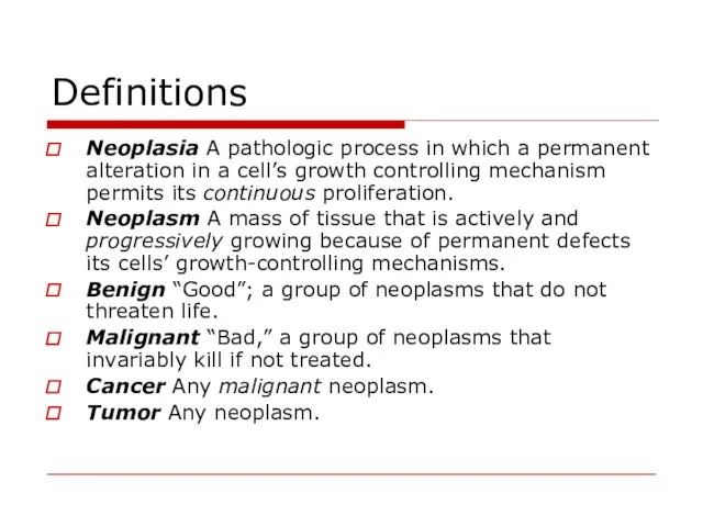
Definitions
Neoplasia A pathologic process in which a permanent alteration in a
Definitions
Neoplasia A pathologic process in which a permanent alteration in a
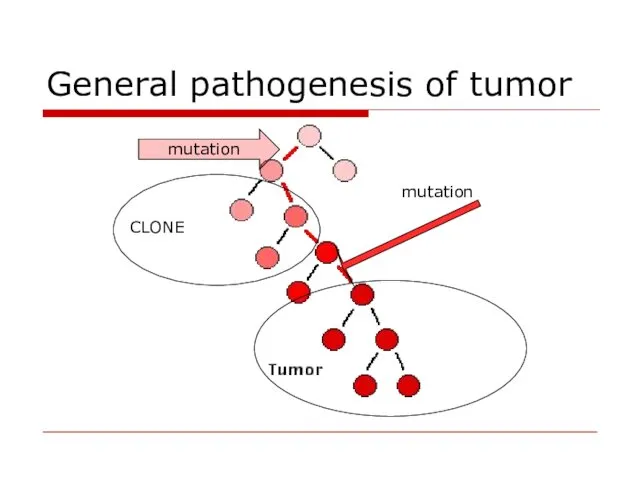
General pathogenesis of tumor
mutation
CLONE
mutation
General pathogenesis of tumor
mutation
CLONE
mutation
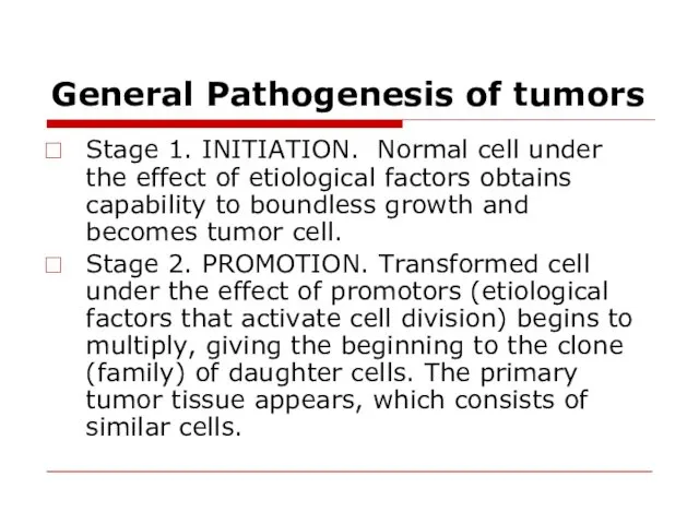
General Pathogenesis of tumors
Stage 1. INITIATION. Normal cell under the
General Pathogenesis of tumors
Stage 1. INITIATION. Normal cell under the
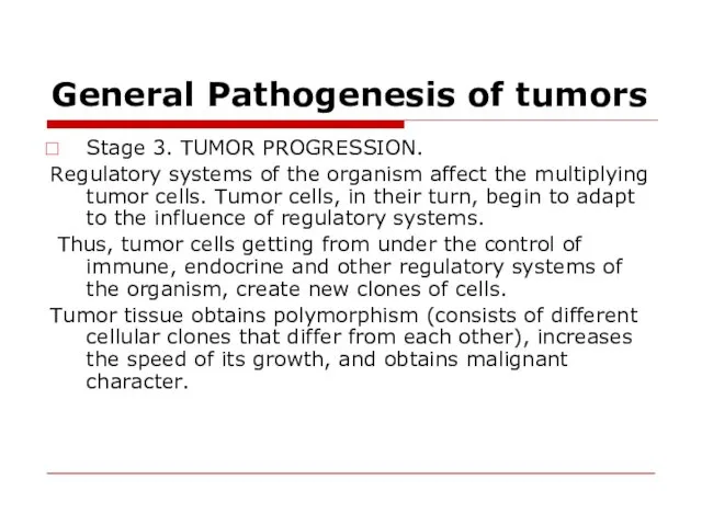
General Pathogenesis of tumors
Stage 3. TUMOR PROGRESSION.
Regulatory systems of the
General Pathogenesis of tumors
Stage 3. TUMOR PROGRESSION.
Regulatory systems of the
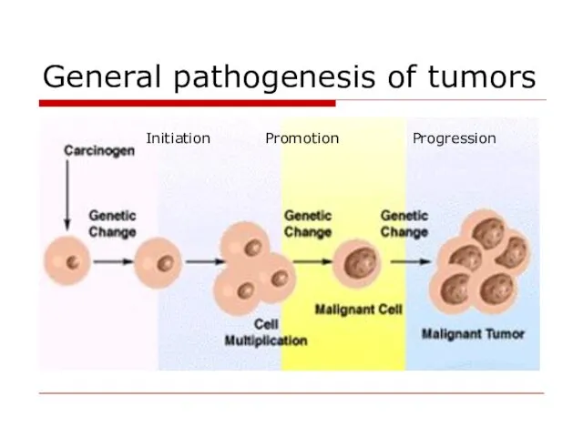
General pathogenesis of tumors
Initiation
Promotion
Progression
General pathogenesis of tumors
Initiation
Promotion
Progression
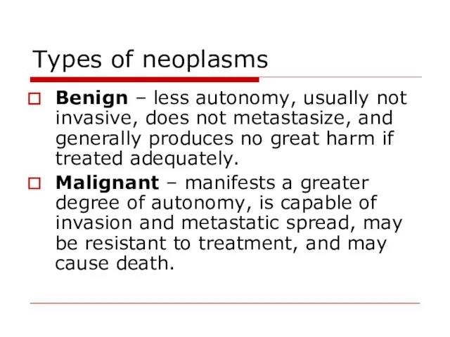
Types of neoplasms
Benign – less autonomy, usually not invasive, does not
Types of neoplasms
Benign – less autonomy, usually not invasive, does not
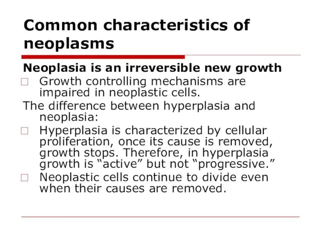
Common characteristics of neoplasms
Neoplasia is an irreversible new growth
Growth
Common characteristics of neoplasms
Neoplasia is an irreversible new growth
Growth
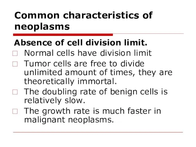
Common characteristics of neoplasms
Absence of cell division limit.
Normal cells have
Common characteristics of neoplasms
Absence of cell division limit.
Normal cells have
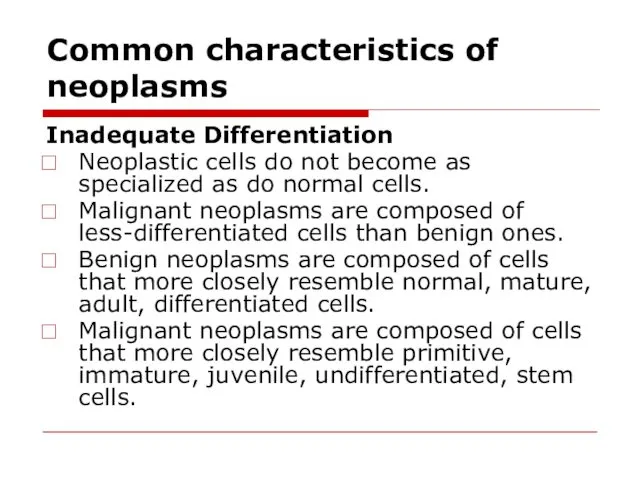
Common characteristics of neoplasms
Inadequate Differentiation
Neoplastic cells do not become as specialized
Common characteristics of neoplasms
Inadequate Differentiation
Neoplastic cells do not become as specialized
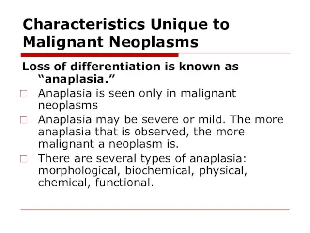
Characteristics Unique to Malignant Neoplasms
Loss of differentiation is known as “anaplasia.”
Anaplasia
Characteristics Unique to Malignant Neoplasms
Loss of differentiation is known as “anaplasia.”
Anaplasia
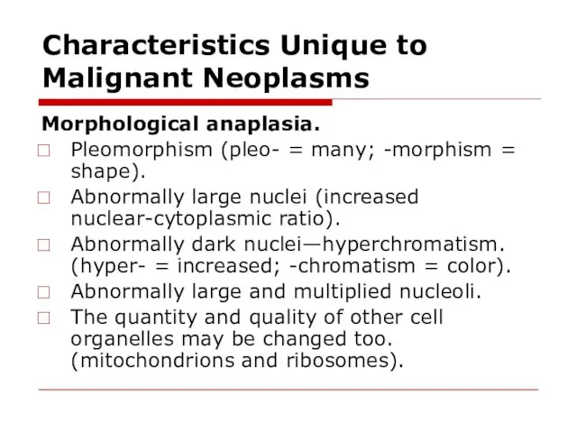
Characteristics Unique to Malignant Neoplasms
Morphological anaplasia.
Pleomorphism (pleo- = many; -morphism
Characteristics Unique to Malignant Neoplasms
Morphological anaplasia.
Pleomorphism (pleo- = many; -morphism
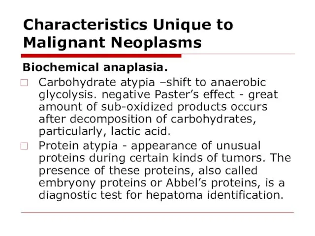
Characteristics Unique to Malignant Neoplasms
Biochemical anaplasia.
Carbohydrate atypia –shift to anaerobic glycolysis.
Characteristics Unique to Malignant Neoplasms
Biochemical anaplasia.
Carbohydrate atypia –shift to anaerobic glycolysis.
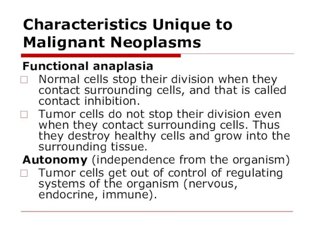
Characteristics Unique to Malignant Neoplasms
Functional anaplasia
Normal cells stop their division
Characteristics Unique to Malignant Neoplasms
Functional anaplasia
Normal cells stop their division
Characteristics Unique to Malignant Neoplasms
Loss of polaruty and specialized functions
Normal differentiated
Characteristics Unique to Malignant Neoplasms
Loss of polaruty and specialized functions
Normal differentiated
Characteristics Unique to Malignant Neoplasms
Invasion
Malignant cells lose their attachment to their
Characteristics Unique to Malignant Neoplasms
Invasion
Malignant cells lose their attachment to their
Characteristics Unique to Malignant Neoplasms
Invasive malignancies are difficult to eradicate.
There is
Characteristics Unique to Malignant Neoplasms
Invasive malignancies are difficult to eradicate.
There is
Characteristics Unique to Malignant Neoplasms
Metastasis
The tendency of malignant neoplasms to spread
Characteristics Unique to Malignant Neoplasms
Metastasis
The tendency of malignant neoplasms to spread
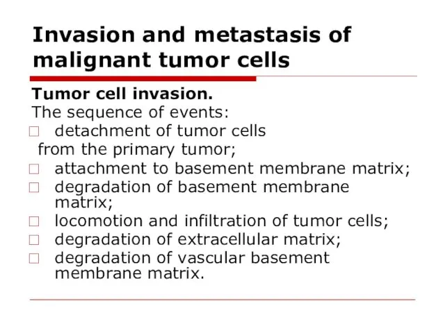
Invasion and metastasis of malignant tumor cells
Tumor cell invasion.
The sequence
Invasion and metastasis of malignant tumor cells
Tumor cell invasion.
The sequence
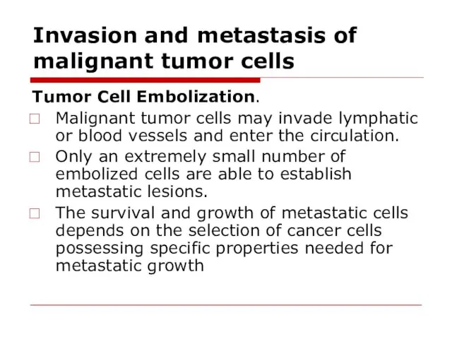
Invasion and metastasis of malignant tumor cells
Tumor Cell Embolization.
Malignant tumor
Invasion and metastasis of malignant tumor cells
Tumor Cell Embolization.
Malignant tumor
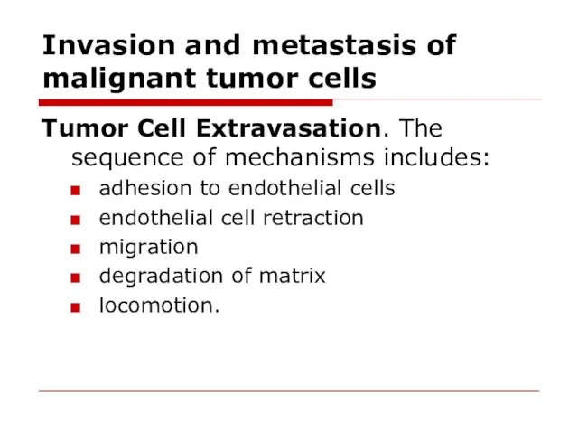
Invasion and metastasis of malignant tumor cells
Tumor Cell Extravasation. The sequence
Invasion and metastasis of malignant tumor cells
Tumor Cell Extravasation. The sequence
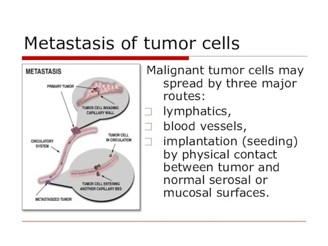
Metastasis of tumor cells
Malignant tumor cells may spread by three major
Metastasis of tumor cells
Malignant tumor cells may spread by three major
Other Differences between Benign and Malignant Neoplasms
Benign Neoplasms growth pattern is
Other Differences between Benign and Malignant Neoplasms
Benign Neoplasms growth pattern is
Other Differences between Benign and Malignant Neoplasms
Malignant neoplasms grow rapidly and
Other Differences between Benign and Malignant Neoplasms
Malignant neoplasms grow rapidly and
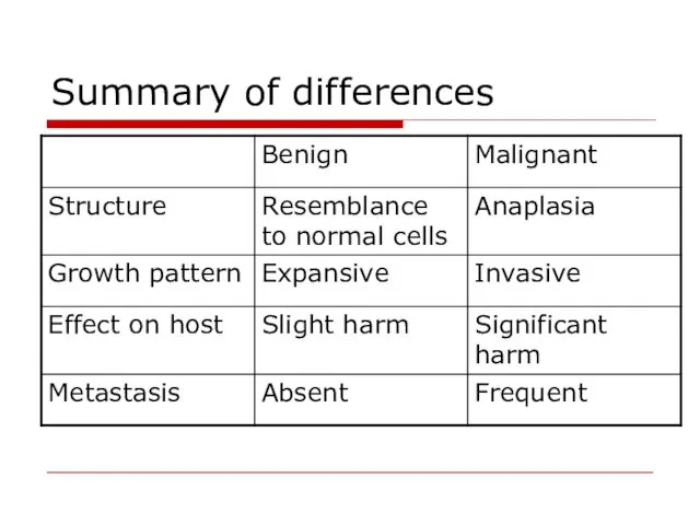
Summary of differences
Summary of differences
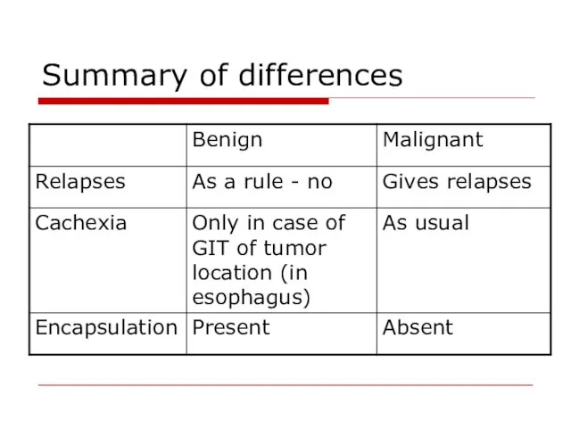
Summary of differences
Summary of differences
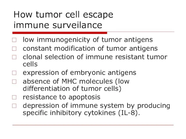
How tumor cell escape
immune surveilance
low immunogenicity of tumor antigens
constant modification
How tumor cell escape
immune surveilance
low immunogenicity of tumor antigens
constant modification
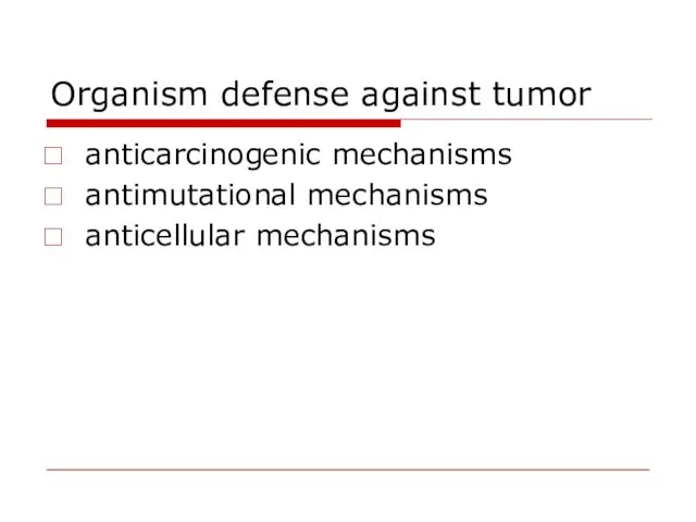
Organism defense against tumor
anticarcinogenic mechanisms
antimutational mechanisms
anticellular mechanisms
Organism defense against tumor
anticarcinogenic mechanisms
antimutational mechanisms
anticellular mechanisms
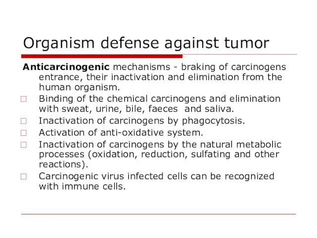
Organism defense against tumor
Anticarcinogenic mechanisms - braking of carcinogens entrance, their
Organism defense against tumor
Anticarcinogenic mechanisms - braking of carcinogens entrance, their
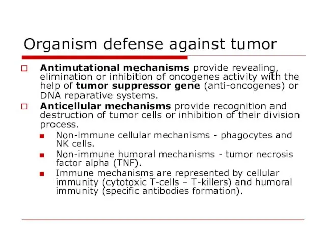
Organism defense against tumor
Antimutational mechanisms provide revealing, elimination or inhibition of
Organism defense against tumor
Antimutational mechanisms provide revealing, elimination or inhibition of
Organism defense against tumor
TNF is released primarily by WBC and endothelium.
Organism defense against tumor
TNF is released primarily by WBC and endothelium.
Interrelations between the host organism and the tumor
Peculiarities of tumor
Location
Interrelations between the host organism and the tumor
Peculiarities of tumor
Location
Interrelations between the host organism and the tumor
Cancer Cachexia is a
Interrelations between the host organism and the tumor
Cancer Cachexia is a
Interrelations between the host organism and the tumor
The paraneoplastic syndromes: hypercalcemia,
Interrelations between the host organism and the tumor
The paraneoplastic syndromes: hypercalcemia,
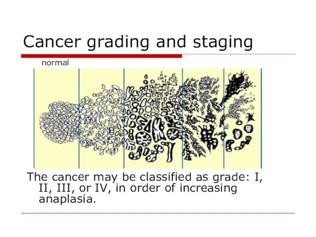
Cancer grading and staging
The cancer may be classified as
Cancer grading and staging
The cancer may be classified as
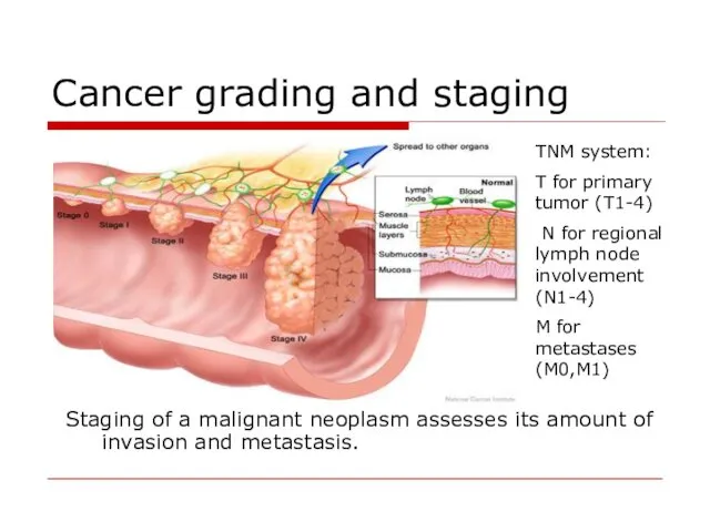
Cancer grading and staging
Staging of a malignant neoplasm assesses its amount
Cancer grading and staging
Staging of a malignant neoplasm assesses its amount
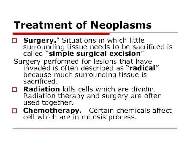
Treatment of Neoplasms
Surgery.” Situations in which little surrounding tissue needs to
Treatment of Neoplasms
Surgery.” Situations in which little surrounding tissue needs to
 Гемолитические анемии
 Определение и гигиеническая оценка режима дня в дошкольном учреждении и расписание уроков в СОШ
 Формирование здорового образа жизни и профилактика хронических неинфекционных заболеваний среди несовершеннолетних
 Повреждение нижних конечностей
 Возможности высокопольной МРТ в диагностике наследственных и редких врожденных заболеваний. Лекция 2
 Анафилактикалық шок
 Биохимия и медицина: предмет, цель, задачи, методы биохимии. Связь биохимии и медицины
 Вміст вуглекислого газу в класній кімнаті та вплив його концентрації на розумову діяльність учнів
 ВИЧ-инфекция и его профилактика
 Гистофизиология органов дыхательной системы
 Общий анализ крови
 Эклампсия
 Боли в спине в практике терапевта и ревматолога
 Иммунодефицитные состояния у детей
 Планування сім′ї
 Көру мүшесінің жарақаттары жайлы түсінік
 Ятрогендік аурулар
 Ортанғы құлақтың созылмалы қабынуы. Бассүйекішілік отогенді асқыну
 Мектепке дайындау
 Обезболивание в хирургии
 Электрокардиограммы у детей в разные возрастные периоды
 Рак пищевода
 Клинические особенности менингококковой инфекции у детей на современном этапе
 Промышленные яды. Токсическое действие на организм человека
 Оценка тяжести состояния пациентов с циррозом печени
 Патофизиология пищеварения
 Денсаулық сақтау жүйесіндегі. Аккредитация
 Трансплантационный иммунитет