Слайд 2
Introduction
In all countries, women above a fixed cut-off age were regarded
as at high enough risk of aneuploidy to warrant the costs and hazards of performing an invasive diagnostic procedure. Over the past three decades, attempts have been made to refine the assessment of an individual woman’s risk using biochemical and ultrasound markers within pregnancy. These have improved the sensitivity (proportion of aneuploidy pregnancies at high risk; or detection rate) and specificity (proportion of unaffected pregnancies not at high risk).3, 4 Using a cut-off maternal age of 35, a 30–40% sensitivity and 90–95% specificity (or 5–10% false-positive rate) were the best available statistics throughout the 1970s and early 1980s.
Слайд 3
First Biochemical Marker
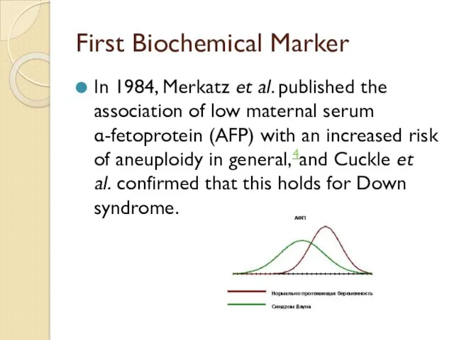
In 1984, Merkatz et al. published the association of low
maternal serum α-fetoprotein (AFP) with an increased risk of aneuploidy in general,4and Cuckle et al. confirmed that this holds for Down syndrome.
Слайд 4
What is it AFP?
it was used to screen for neural
tube defects, at 16–18 weeks' gestation, it was relatively simple to extend the test interpretation to include aneuploidy.
This was done by the calculation of a likelihood ratio (proportion of aneuploidy pregnancies divided by proportion of unaffected pregnancies with the given AFP level) and using this to increase or decrease the maternal age-specific risk.
Слайд 5
A brief history of AFP
Maternal serum AFP screening for aneuploidy was
widely adopted and had the potential to increase the detection rate, but it was inefficient. The optimal use of a biochemical or ultrasound marker is to screen all women regardless of age and to define high risk purely on the basis of the screening result. However, many clinicians did not consider a low risk AFP result in an older woman as sufficient grounds for not offering invasive testing. While the use of maternal serum AFP was a notable improvement over “how old are you?”, it left much to be desied.
Слайд 6
FIRST HIGHLY DISCRIMINATORY MARKER
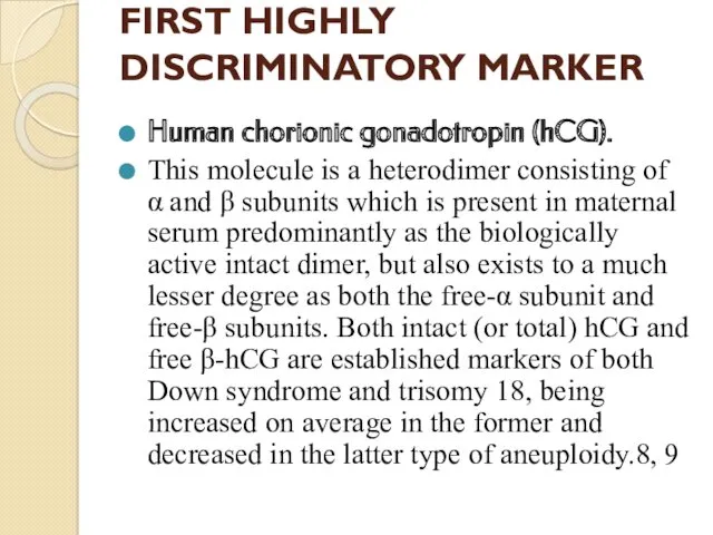
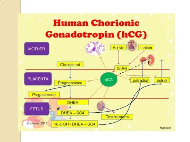
Human chorionic gonadotropin (hCG).
This molecule is a heterodimer
consisting of α and β subunits which is present in maternal serum predominantly as the biologically active intact dimer, but also exists to a much lesser degree as both the free-α subunit and free-β subunits. Both intact (or total) hCG and free β-hCG are established markers of both Down syndrome and trisomy 18, being increased on average in the former and decreased in the latter type of aneuploidy.8, 9
Слайд 7
Слайд 8
Power of uE3
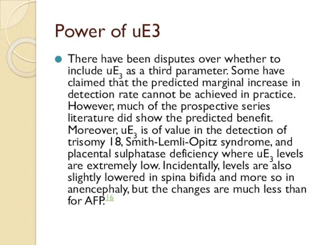
There have been disputes over whether to include uE3 as
a third parameter. Some have claimed that the predicted marginal increase in detection rate cannot be achieved in practice. However, much of the prospective series literature did show the predicted benefit. Moreover, uE3 is of value in the detection of trisomy 18, Smith-Lemli-Opitz syndrome, and placental sulphatase deficiency where uE3 levels are extremely low. Incidentally, levels are also slightly lowered in spina bifida and more so in anencephaly, but the changes are much less than for AFP.16
Слайд 9
MULTIPLE BIOCHEMICAL MARKERS
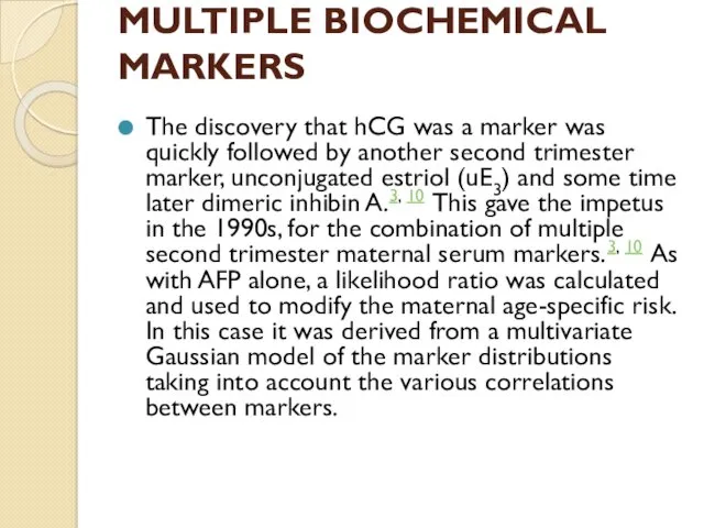
The discovery that hCG was a marker was quickly
followed by another second trimester marker, unconjugated estriol (uE3) and some time later dimeric inhibin A.3, 10 This gave the impetus in the 1990s, for the combination of multiple second trimester maternal serum markers.3, 10 As with AFP alone, a likelihood ratio was calculated and used to modify the maternal age-specific risk. In this case it was derived from a multivariate Gaussian model of the marker distributions taking into account the various correlations between markers.
Слайд 10
Слайд 11
Another long promising but yet to be fulfilled marker was the
search for fetal cells in maternal circulation. Studies throughout the 1990s and early 2000s suggested that isolation and analysis of fetal cells might, in fact, become practical and useful as a screening test.17, 18 Much of the 1980s and 1990s focused on ways to improve the efficacy of detection methods primarily centered on the need to increase the enrichment of fetal cells from the maternal blood circulation the prevalence of which has been estimated to be approximately 1 in 10,000,000 cells with no clear likelihood of success.19After the failure of the first lines of methodology in detecting fetal cells, modified approaches have emerged that are being evaluated for more precise identification and isolation of fetal cells.
Слайд 12
SEQUENTIAL SCREENING METHODS
Three types of sequential policy have received attention. The
first to be proposed was a form of non-disclosure sequential screening using first trimester PAPP-A and NT together with second trimester AFP, uE3, free β-hCG or intact hCG, and inhibin (integrated test). Risks are not used clinically until all markers have been tested. The proponents of such “integrated” screening argue that higher sensitivities can be achieved and therefore justify the nondisclosure. However, many clinicians in the United States and elsewhere feel that it is simply not acceptable under local culture and ethical beliefs to withhold potentially serious screening results for a month when the odds of substantial change are minimal. Such an approach also has the substantial disadvantage that there is no early diagnosis or reassurance.
Слайд 13
A second approach (step-wise test) begins with first trimester PAPP-A, free β-hCG
or intact hCG, and NT; those with low risk have second trimester AFP, uE3, free β-hCG or intact hCG, and inhibin; the risk is estimated from all seven markers. It is important to use a higher first trimester cut-off than with non-sequential screening, otherwise the overall false-positive rate will be too high. And it is essential to use all seven markers together when calculating the final risk. It is invalid to ignore the first trimester markers at this stage although many practitioners are doing so because they do not have access to the appropriate risk calculation software. This policy restores some first trimester diagnosis.
A third policy, more efficient than the other types, is called the contingent test. This begins with first trimester PAPP-A, free β-hCG or intact hCG, and NT. Women with very high risk are offered immediate invasive prenatal diagnosis and only those with borderline risks are offered second trimester AFP, uE3, free β-hCG or intact hCG, and inhibin; their risk is estimated from all seven markers. The borderline is chosen so that a large proportion of women have early assurance. This group has such a low risk that it is very unlikely that further markers will lead to a final high risk result.
Слайд 14
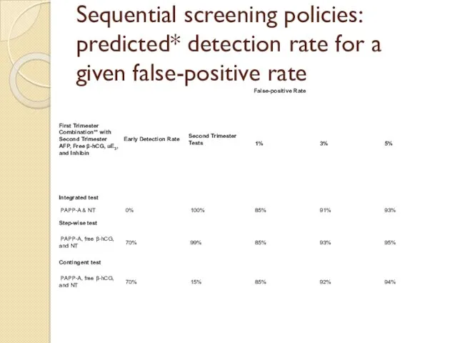
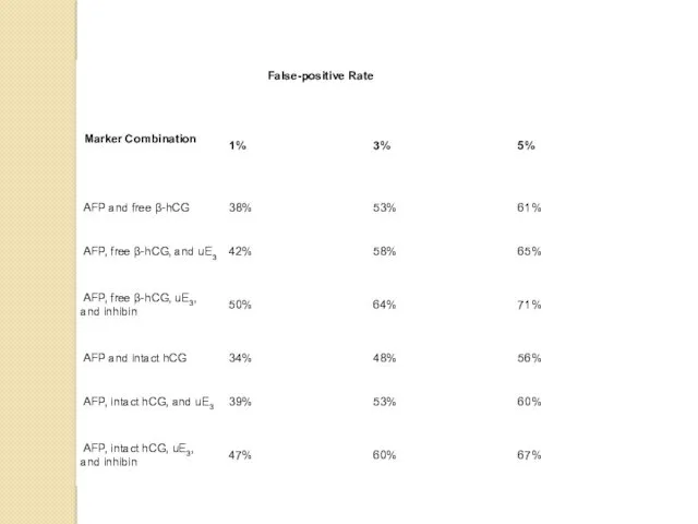
Sequential screening policies: predicted* detection rate for a given false-positive rate

 Автоматизации твердого звука [ Р] в словах со стечением согласных
 Газовая сварка стали
 Визуальные словари для изучения английского языка
 Презентация по проекту.pptx
 Исследовательская работа по краеведению Топонимы Белогорского района Амурской области
 Введение в зоологию. Подцарство Одноклеточные или Простейшие (Protozoa)
 Игры для адаптации детей младшего возраста в период логопедического мониторинга.
 Презентация к у року в 6 классе на тему Атмосферные осадки
 СССР в последние десятилетия советской власти (1965 - 1991)
 04 (2)
 Расчет короба под динамик DD3512H
 Дорожный фонд Республики Беларусь
 Совершенствование коррекционно-развивающей среды логопедического кабинета в условиях реализации ФГОС
 Oil industry Jargon
 Биохимия крови
 Технолігїї підвищення продуктивності процесорів
 Презентация Развитие творческих качеств педагога как условие развития личностных качеств и творческих способностей дошкольников
 Питание в туристском походе
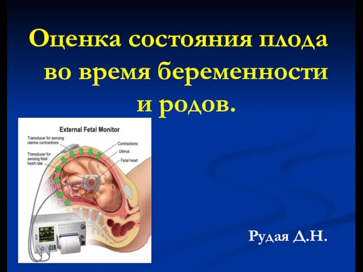 Оценка состояния плода во время беременности и родов
 История Казахстана
 Нормативно-правовые акты по охране труда
 Прославлення і хвала
 лягушонок _ cvc words _ by Artem Morozov
 Викторина: Цветы
 Общение как социально-психологическая категория
 Экономическая политика Петра I
 Ю.А.Гагарин и отряд первых космонавтов на Саратовской земле
 Педагогический совет.