- Introduction & overview

Содержание
- 2. Олег Борисович Птицын (1929-1999)
- 3. PROTEIN PHYSICS LECTURE 1 Introduction & overview
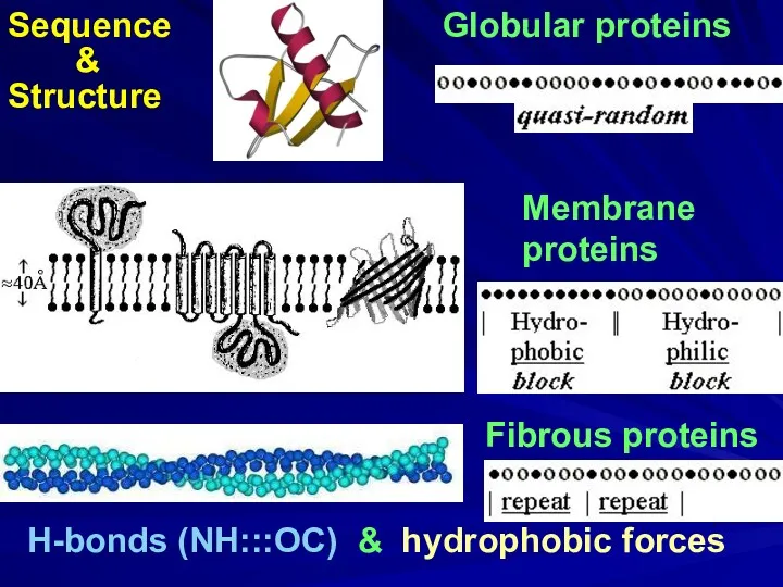
- 4. Globular proteins Fibrous proteins H-bonds (NH:::OC) & hydrophobic forces Membrane proteins
- 5. Protein chain (gene-encoded sequence)
- 6. Secondary structures (α-helices, β-strands) are most conserved structural elements. They form a basis of protein classification
- 9. Globular proteins Fibrous proteins H-bonds (NH:::OC) & hydrophobic forces Membrane proteins Sequence & Structure
- 10. Globular domains C A T H
- 11. PROTEIN CHAIN CAN FORM ITS UNIQUE 3D STRUCTURE SPONTANEOUSLY IN VITRO
- 12. phase separation
- 13. BIND ? TRANSFORM ? RELEASE: ENZYMES (chymotrypsin) Note small active site
- 14. POST-TRANSLATIONAL MODIFICATIONS Sometimes, CHAIN CUT-INDUCED DEFORMATION MAKES ENZYME ACTIVE Chymotripsin Chymotripsinogen active cat. site non-active cat.
- 15. POST-TRANSLATIONAL MODIFICATIONS: (especially in eukaryotes): PROTEIN CHAIN CUTS (proteolysis), - SPLICING (inteins) - CYCLIZATION - INTERNAL
- 16. Sometimes: Different folds with the same active site: the same biochemical function
- 17. Sometimes: Similar folds with different active sites: different biochemical function 4-helix bundle COFACTORS: HEME, 2Fe, RNA,
- 18. Standard positions of active sites in protein folds
- 19. Natively disordered protein: X-ray + SAXS + NMR + MD simulations
- 20. Chaperone GroEL
- 21. ______ NMR
- 22. Protein engineering Wanted: new protein with additional salt bridge (e.g., His+:::Asp-)
- 23. PROTEIN PHYSICS LECTURE 2 Elementary interactions: covalent
- 24. Protein chain: regular backbone & gene-encoded sequence of side chains
- 25. Protein chain Covalent bond lengths: 0.9 – 1.8 Å Covalent bond angles: 109o – 120o Atom
- 26. Side chains
- 27. Main-chain: peptide group: flat & rigid Side chains: L amino acids ___ ______ ______ Protein chain
- 28. Ala _L Gly Thr Ile Two asymmetric side chains: Symmetric Asymmetric backbone-to- side_chain: Stereo images
- 29. ~ V = ±|V| ≅ semi-classical approximation
- 30. Werner Karl Heisenberg (1901-76) — Nobel Prize 1932 Wolfgang Ernst Pauli ) (1900-58) — Nobel Prize
- 31. Peptide group: flat & rigid sp2 + p sp2 + p Covalent bonding in peptide group:
- 32. Main-chain: φ (N-Cα) , ψ (Cα-C’), ω (C’=N) Side-chain: χ1, χ2, ...
- 33. Counting angles: _____________________________________________ 0o 180o 120o
- 34. sp2 - sp2 (ω) ω = 180o ω = 0o
- 35. Potentials: from IR spectra of vibrations sp2 - sp2 (ω) sp3 – sp3 (χ) sp2 –
- 36. Harold Abraham Scheraga (1921) Paul John Flory (1910-85) — Nobel Prize 1974 Александр Исаакович Китайгородский (1914–1985)
- 38. Скачать презентацию
Олег Борисович Птицын
(1929-1999)
Олег Борисович Птицын
(1929-1999)
PROTEIN PHYSICS
LECTURE 1
Introduction & overview
PROTEIN PHYSICS
LECTURE 1
Introduction & overview
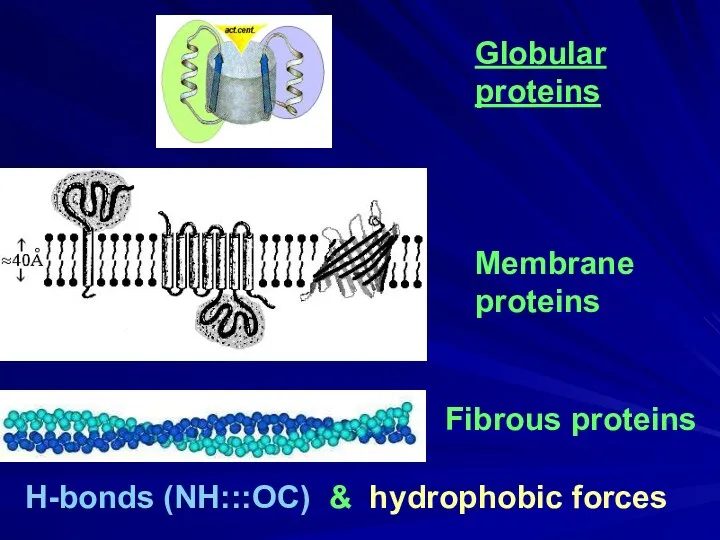
Globular
proteins
Fibrous proteins
H-bonds (NH:::OC) & hydrophobic forces
Membrane
proteins
Globular
proteins
Fibrous proteins
H-bonds (NH:::OC) & hydrophobic forces
Membrane
proteins
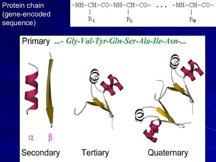
Protein chain
(gene-encoded sequence)
Protein chain
(gene-encoded sequence)
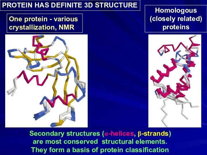
Secondary structures (α-helices, β-strands)
are most conserved structural elements.
They form
Secondary structures (α-helices, β-strands)
are most conserved structural elements.
They form
Globular proteins
Fibrous proteins
H-bonds (NH:::OC) & hydrophobic forces
Membrane
proteins
Sequence
&
Structure
Globular proteins
Fibrous proteins
H-bonds (NH:::OC) & hydrophobic forces
Membrane
proteins
Sequence
&
Structure
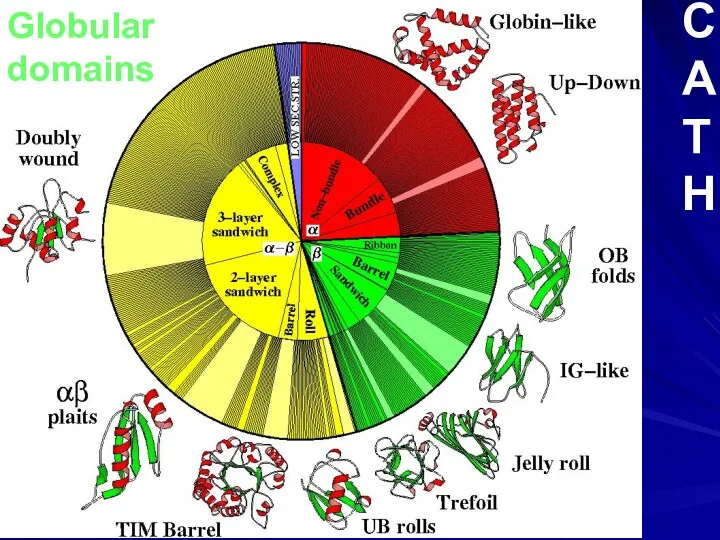
Globular
domains
C
A
T
H
Globular
domains
C
A
T
H
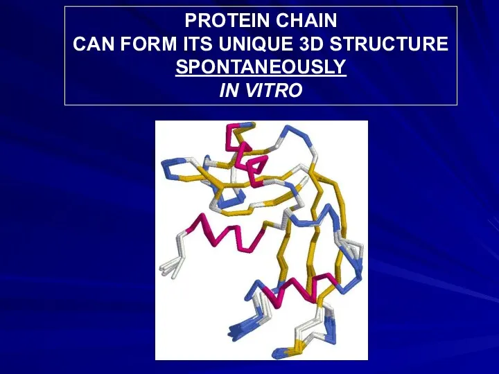
PROTEIN CHAIN
CAN FORM ITS UNIQUE 3D STRUCTURE
SPONTANEOUSLY
IN VITRO
PROTEIN CHAIN
CAN FORM ITS UNIQUE 3D STRUCTURE
SPONTANEOUSLY
IN VITRO
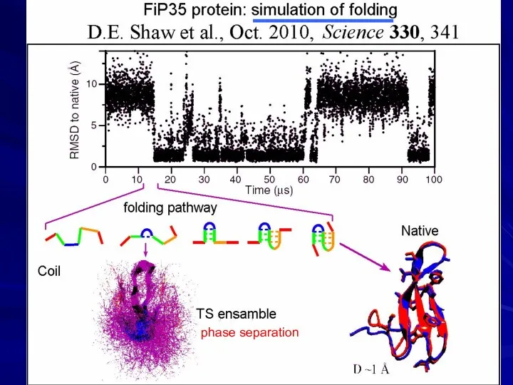
phase separation
phase separation
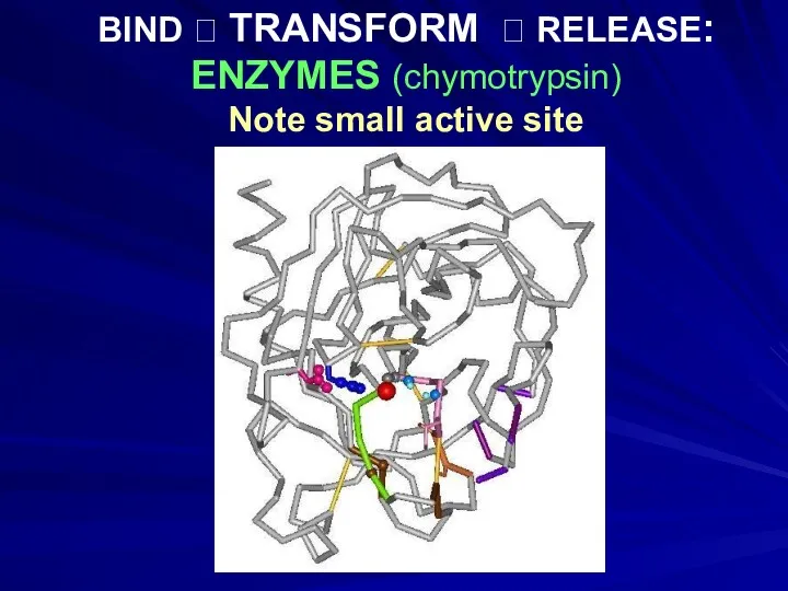
BIND ? TRANSFORM ? RELEASE:
ENZYMES (chymotrypsin)
Note small active site
BIND ? TRANSFORM ? RELEASE:
ENZYMES (chymotrypsin)
Note small active site
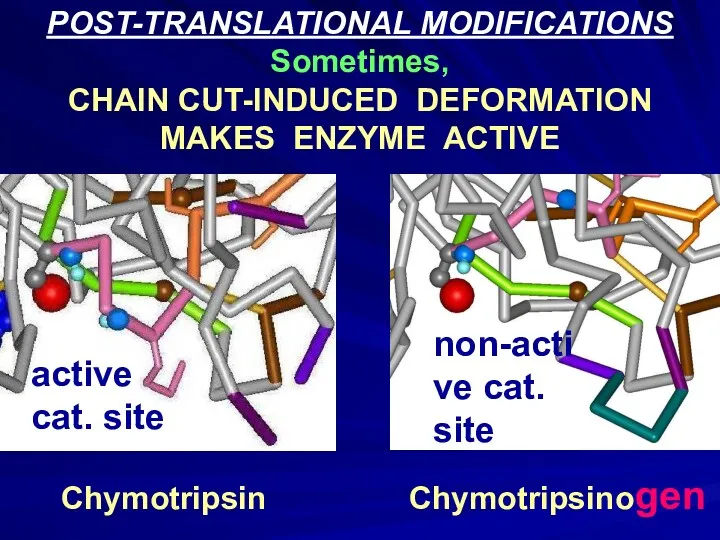
POST-TRANSLATIONAL MODIFICATIONS
Sometimes,
CHAIN CUT-INDUCED DEFORMATION MAKES ENZYME ACTIVE
Chymotripsin Chymotripsinogen
active cat. site
non-active cat.
POST-TRANSLATIONAL MODIFICATIONS
Sometimes,
CHAIN CUT-INDUCED DEFORMATION MAKES ENZYME ACTIVE
Chymotripsin Chymotripsinogen
active cat. site
non-active cat.
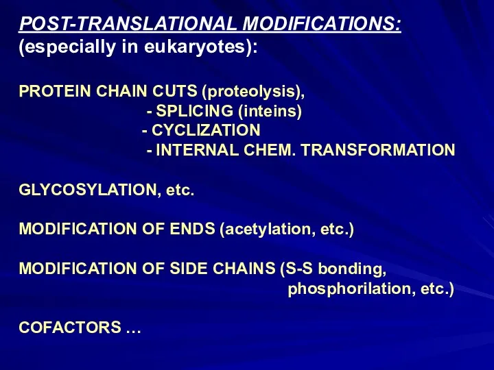
POST-TRANSLATIONAL MODIFICATIONS:
(especially in eukaryotes):
PROTEIN CHAIN CUTS (proteolysis),
- SPLICING (inteins) -
POST-TRANSLATIONAL MODIFICATIONS: (especially in eukaryotes): PROTEIN CHAIN CUTS (proteolysis), - SPLICING (inteins) -
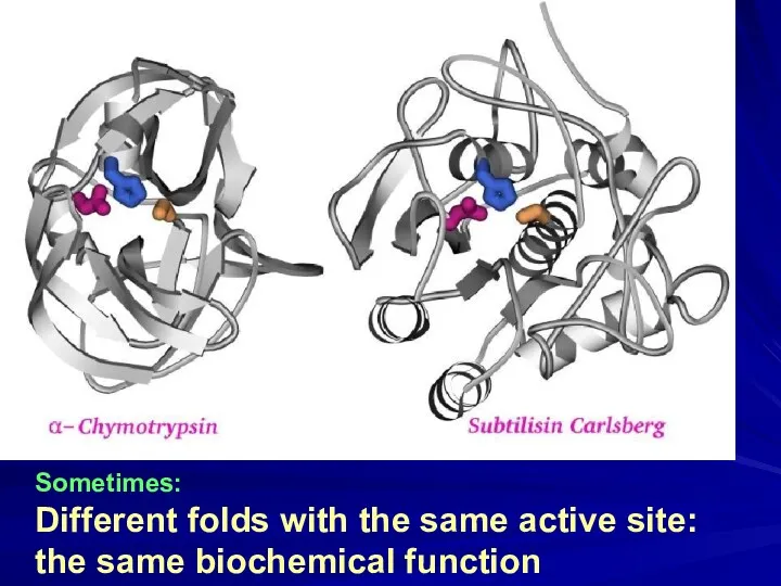
Sometimes:
Different folds with the same active site:
the same biochemical function
Sometimes:
Different folds with the same active site:
the same biochemical function
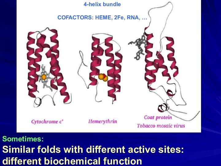
Sometimes:
Similar folds with different active sites: different biochemical function
4-helix bundle
COFACTORS:
Sometimes:
Similar folds with different active sites: different biochemical function
4-helix bundle
COFACTORS:
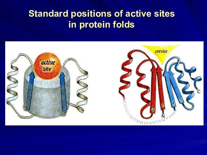
Standard positions of active sites in protein folds
Standard positions of active sites in protein folds
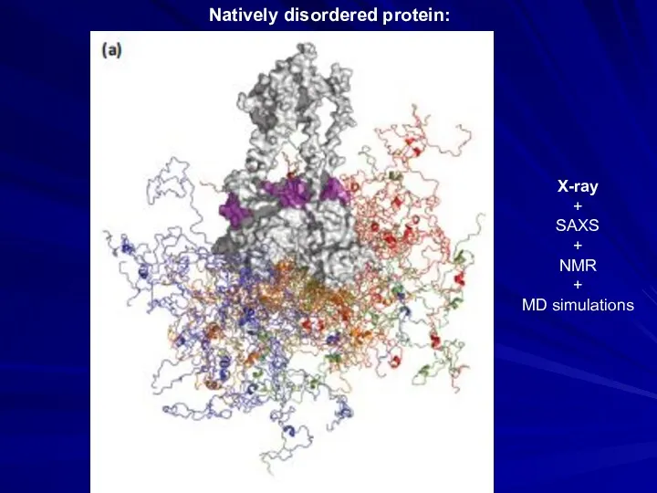
Natively disordered protein:
X-ray
+
SAXS
+
NMR
+
MD simulations
Natively disordered protein:
X-ray
+
SAXS
+
NMR
+
MD simulations
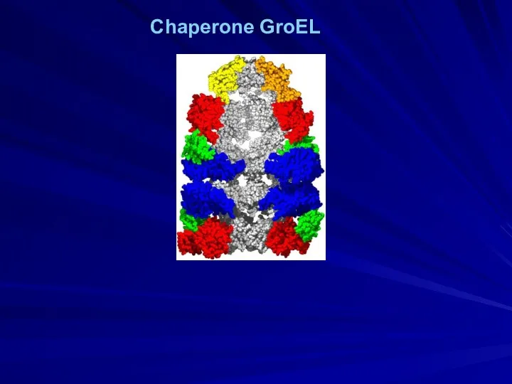
Chaperone GroEL
Chaperone GroEL
______
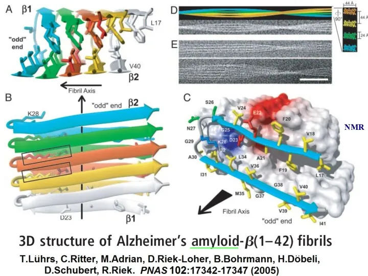
NMR
______
NMR
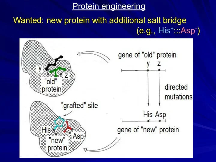
Protein engineering
Wanted: new protein with additional salt bridge
(e.g., His+:::Asp-)
Protein engineering
Wanted: new protein with additional salt bridge
(e.g., His+:::Asp-)
PROTEIN PHYSICS
LECTURE 2
Elementary interactions:
covalent
PROTEIN PHYSICS
LECTURE 2
Elementary interactions:
covalent
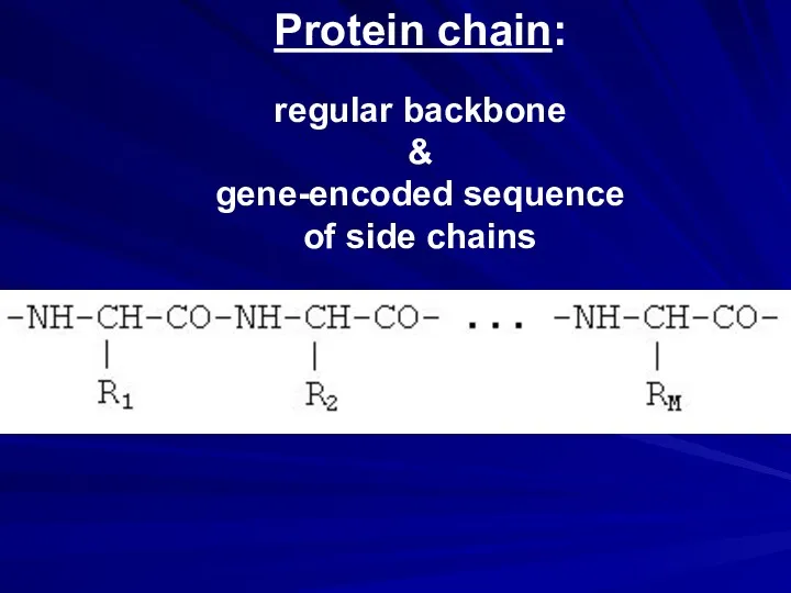
Protein chain:
regular backbone
&
gene-encoded sequence
of side chains
Protein chain:
regular backbone
&
gene-encoded sequence
of side chains
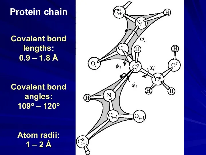
Protein chain
Covalent bond lengths:
0.9 – 1.8 Å
Covalent bond angles:
109o – 120o
Atom
Protein chain
Covalent bond lengths:
0.9 – 1.8 Å
Covalent bond angles:
109o – 120o
Atom
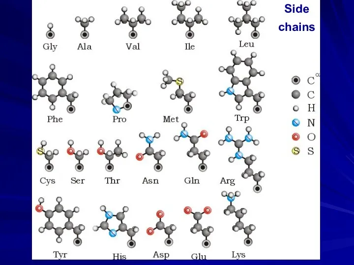
Side chains
Side chains
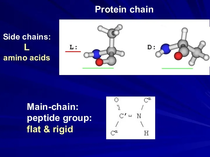
Main-chain:
peptide group:
flat & rigid
Side chains: L
amino acids
___
______
______
Protein chain
Main-chain:
peptide group:
flat & rigid
Side chains: L
amino acids
___
______
______
Protein chain
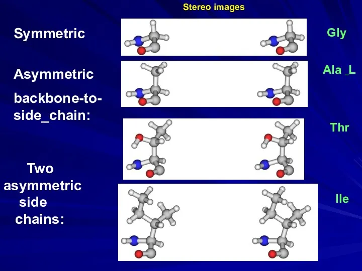
Ala _L
Gly
Thr
Ile
Two
asymmetric
side
chains:
Symmetric
Asymmetric
backbone-to- side_chain:
Stereo images
Ala _L
Gly
Thr
Ile
Two
asymmetric
side
chains:
Symmetric
Asymmetric
backbone-to- side_chain:
Stereo images
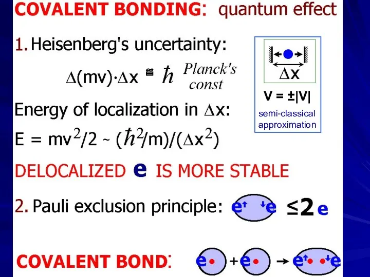
~
V = ±|V|
≅
semi-classical
approximation
~
V = ±|V|
≅
semi-classical
approximation
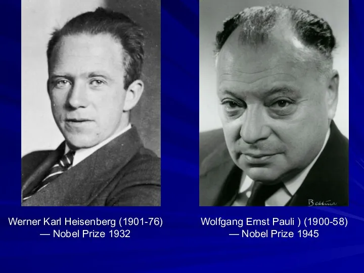
Werner Karl Heisenberg (1901-76)
— Nobel Prize 1932
Wolfgang Ernst Pauli ) (1900-58)
— Nobel Prize 1945
Werner Karl Heisenberg (1901-76)
— Nobel Prize 1932
Wolfgang Ernst Pauli ) (1900-58)
— Nobel Prize 1945
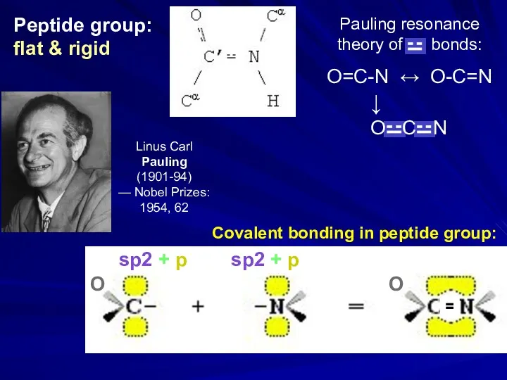
Peptide group:
flat & rigid
sp2 + p sp2 + p
Covalent bonding
Peptide group:
flat & rigid
sp2 + p sp2 + p
Covalent bonding
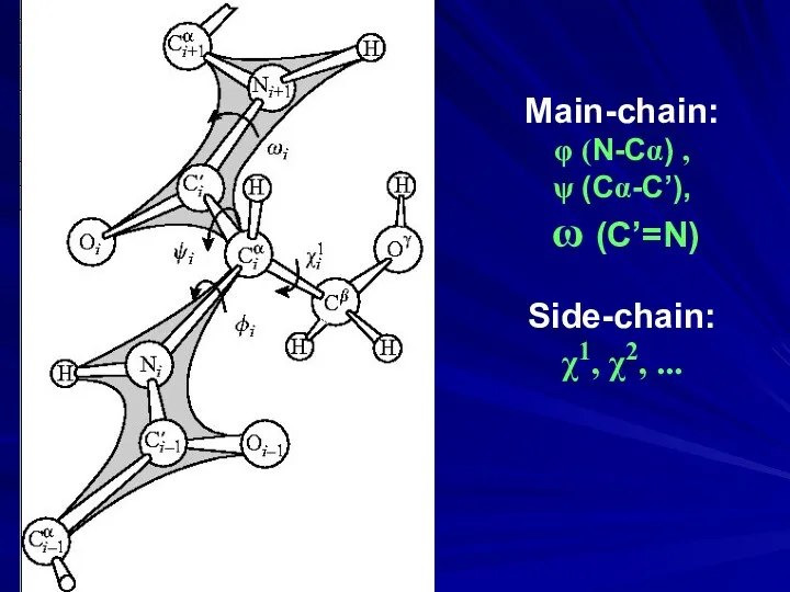
Main-chain:
φ (N-Cα) ,
ψ (Cα-C’),
ω (C’=N)
Side-chain:
χ1, χ2, ...
Main-chain:
φ (N-Cα) ,
ψ (Cα-C’),
ω (C’=N)
Side-chain:
χ1, χ2, ...
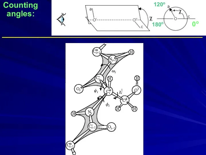
Counting
angles:
_____________________________________________
0o
180o
120o
Counting
angles:
_____________________________________________
0o
180o
120o
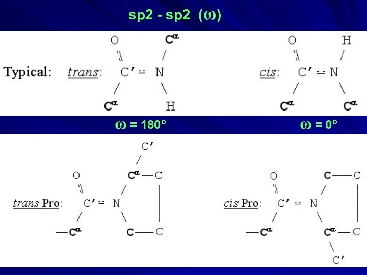
sp2 - sp2 (ω)
ω = 180o ω = 0o
sp2 - sp2 (ω)
ω = 180o ω = 0o
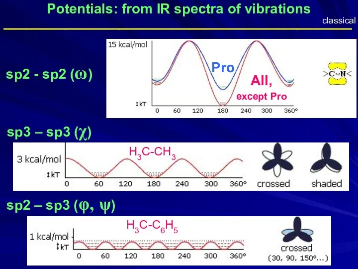
Potentials: from IR spectra of vibrations
sp2 - sp2 (ω)
sp3
Potentials: from IR spectra of vibrations
sp2 - sp2 (ω)
sp3
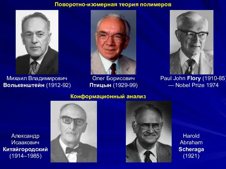
Harold
Abraham
Scheraga
(1921)
Paul John Flory (1910-85)
— Nobel Prize 1974
Александр
Исаакович
Китайгородский
(1914–1985)
Михаил Владимирович
Волькенштейн (1912-92)
Олег Борисович
Птицын
Harold
Abraham
Scheraga
(1921)
Paul John Flory (1910-85)
— Nobel Prize 1974
Александр
Исаакович
Китайгородский
(1914–1985)
Михаил Владимирович
Волькенштейн (1912-92)
Олег Борисович
Птицын
 Конструкция и технология изготовления каркаса руля высоты самолета АН-148
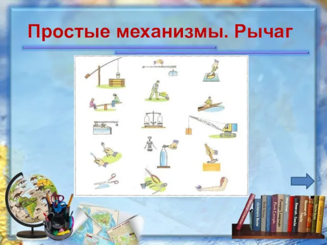 Простые механизмы. Рычаг
 Электр өрісі
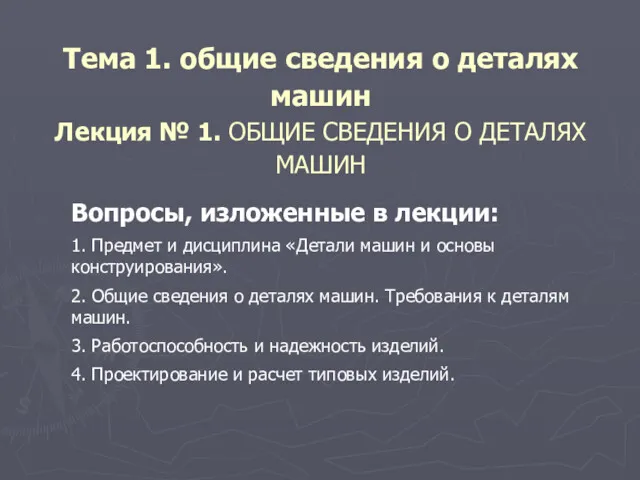 Общие сведения о деталях машин
 к уроку физики в 7 классе на тему: Простые механизмы в картинках и задачи в картинках
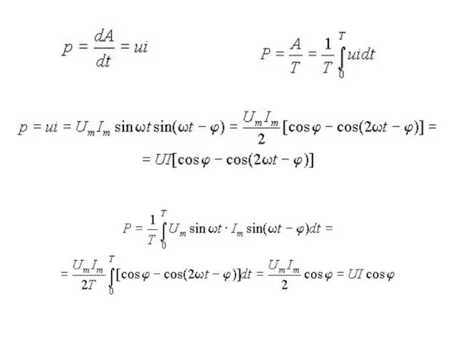 Компенсация реактивной мощности. Потребители реактивной мощности. Лекция 01
 Механическая энергия. Кинетическая и потенциальная энергия. Закон сохранения энергии
 Термоэлектрические и термомагнитные явления. Диффузионный ток
 Диагностирование системы охлаждения двигателя
 Полевые транзисторы. Классификация полевых транзисторов. Лекция 9
 Оливин. Физические и химические свойства
 Поле в диэлектриках
 Привод устройства сцепления автомобиля Камаз
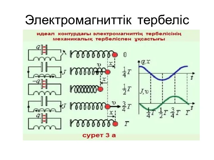 Электромагниттік тербеліс
 Электродинамика
 Электропроводность диэлектриков
 Классификация магнитных методов контроля
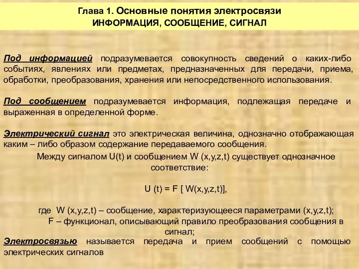 Основные понятия электросвязи. Информация, сообщение, сигнал
 Электротехника и электроника. Электрические машины
 Электрическое сопротивление проводников
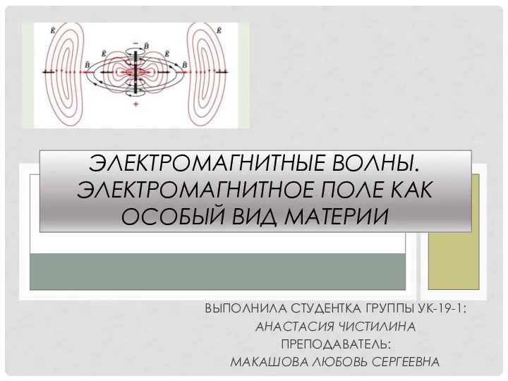 Электромагнитные волны. Электромагнитное поле как особый вид материи
 Гармонические колебания и их характеристики
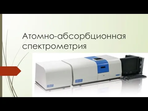 Атомно-абсорбционная спектрометрия
 Электроборудование пассажирских вагонов
 Mass spectrometry
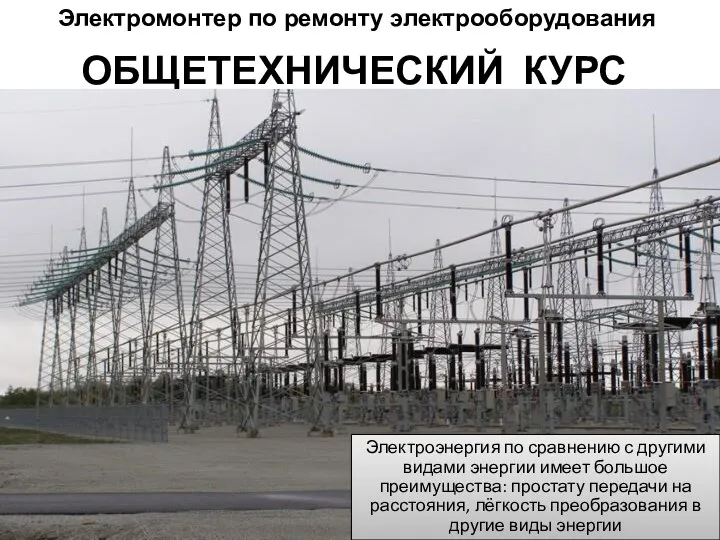 Электромонтер по ремонту электрооборудования
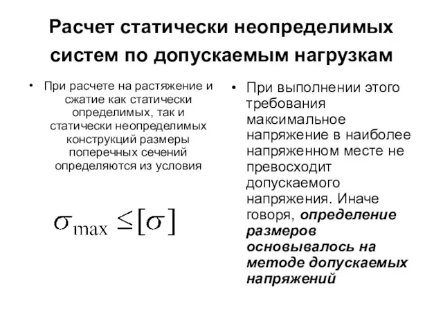 Расчет статически неопределимых систем по допускаемым нагрузкам
 Применение электромагнитных волн