- Anatomy & physiology. The tissue level of organization

Содержание
- 2. FIGURE 4.1 Micrograph of Cervical Tissue This figure is a view of the regular architecture of
- 3. MAJOR CHAPTER OBJECTIVES Identify the main tissue types and discuss their roles in the human body
- 4. 4.1 TYPES OF TISSUES MAJOR SECTION OBJECTIVES Identify the four main tissue types Discuss the functions
- 5. FIGURE 4.2 Four Types of Tissue: Body The four types of tissues are exemplified in nervous
- 6. FIGURE 4.3 Embryonic Origin of Tissues and Major Organs
- 7. FIGURE 4.4 Tissue Membranes The two broad categories of tissue membranes in the body are (1)
- 8. 4.2 EPITHELIAL TISSUE MAJOR SECTION OBJECTIVES Explain the structure and function of epithelial tissue Distinguish between
- 9. FIGURE 4.5 Types of Cell Junctions The three basic types of cell-to-cell junctions are tight junctions,
- 10. FIGURE 4.6 Cells of Epithelial Tissue Simple epithelial tissue is organized as a single layer of
- 11. FIGURE 4.7 Goblet Cell In the lining of the small intestine, columnar epithelium cells are interspersed
- 12. FIGURE 4.8A Summary of Epithelial Tissue Cells
- 13. FIGURE 4.8B Summary of Epithelial Tissue Cells
- 14. FIGURE 4.9 Types of Exocrine Glands Exocrine glands are classified by their structure.
- 15. FIGURE 4.10 Modes of Glandular Secretion In merocrine secretion, the cell remains intact. – E.g. as
- 16. FIGURE 4.11 Sebaceous Glands These glands secrete oils that lubricate and protect the skin. They are
- 17. TABLE 4.1 Connective Tissues Examples
- 18. 4.3 CONNECTIVE TISSUE SUPPORTS AND PROTECTS MAJOR SECTION OBJECTIVES Identify and distinguish between the various sub-types
- 19. FIGURE 4.12 Connective Tissue Proper Fibroblasts produce this fibrous tissue. Connective tissue proper includes the fixed
- 20. FIGURE 4.13 Adipose Tissue This is a loose connective tissue that consists of fat cells with
- 21. FIGURE 4.14 Reticular Tissue This is a loose connective tissue made up of a network of
- 22. FIGURE 4.15 Dense Connective Tissue Dense regular connective tissue consists of collagenous fibers packed into parallel
- 23. FIGURE 4.16 Types of Cartilage Cartilage is a connective tissue consisting of collagenous fibers embedded in
- 24. FIGURE 4.17 Blood: A Fluid Connective Tissue Blood is a fluid connective tissue containing erythrocytes and
- 25. 4.4 MUSCLE TISSUE AND MOTION MAJOR SECTION OBJECTIVES Identify the three types of muscle tissue Compare
- 26. TABLE 4.2 Comparison of Structure and Properties of Muscle Tissue Types
- 27. FIGURE 4.18 Muscle Tissue Skeletal muscle cells have prominent striations and multiple nuclei on their periphery.
- 28. BETTER MUSCLE TISSUE SLIDES Image source: https://www.slideshare.net/syedshahzaib1/lecture-10-muscle-histology accessed 05/26/2017
- 29. 4.5 NERVOUS TISSUE FOR PERCEPTION, RESPONSE MAJOR SECTION OBJECTIVES Identify the classes of cells that make
- 30. FIGURE 4.19 The Neuron The cell body of a neuron, also called the soma, contains the
- 31. FIGURE 4.20 Nervous Tissue Nervous tissue is made up of neurons and neuroglia. The cells of
- 32. 4.6 TISSUE INJURY AND AGING MAJOR SECTION OBJECTIVES Identify the cardinal signs of inflammation List the
- 33. FIGURE 4.21 Tissue Healing During wound repair, collagen fibers are laid down randomly by fibroblasts that
- 34. FIGURE 4.22 Development of Cancer Note the change in cell size, nucleus size, and organization in
- 35. DISORDERS & HOMEOSTATIC IMBALANCES Connective Tissue: Tendinitis Tissues and Cancer Cancer is a generic term for
- 36. INTERACTIVE LINKS View this slideshow http://openstaxcollege.org/l/stemcells about stem cells. View the University of MichiganWebScope at http://openstaxcollege.org/l/goblet
- 38. Скачать презентацию

FIGURE 4.1
Micrograph of Cervical Tissue
This figure is a view of the
FIGURE 4.1
Micrograph of Cervical Tissue
This figure is a view of the

MAJOR CHAPTER OBJECTIVES
Identify the main tissue types and discuss their roles
MAJOR CHAPTER OBJECTIVES
Identify the main tissue types and discuss their roles

4.1 TYPES OF TISSUES
MAJOR SECTION OBJECTIVES
Identify the four main tissue types
Discuss
4.1 TYPES OF TISSUES
MAJOR SECTION OBJECTIVES
Identify the four main tissue types
Discuss

FIGURE 4.2
Four Types of Tissue: Body
The four types of tissues are
FIGURE 4.2
Four Types of Tissue: Body
The four types of tissues are

FIGURE 4.3
Embryonic Origin of Tissues and Major Organs
FIGURE 4.3
Embryonic Origin of Tissues and Major Organs

FIGURE 4.4
Tissue Membranes
The two broad categories of tissue membranes in the
FIGURE 4.4
Tissue Membranes
The two broad categories of tissue membranes in the

4.2 EPITHELIAL TISSUE
MAJOR SECTION OBJECTIVES
Explain the structure and function of epithelial
4.2 EPITHELIAL TISSUE
MAJOR SECTION OBJECTIVES
Explain the structure and function of epithelial

FIGURE 4.5
Types of Cell Junctions
The three basic types of cell-to-cell junctions
FIGURE 4.5
Types of Cell Junctions
The three basic types of cell-to-cell junctions

FIGURE 4.6
Cells of Epithelial Tissue
Simple epithelial tissue is organized as a
FIGURE 4.6
Cells of Epithelial Tissue
Simple epithelial tissue is organized as a

FIGURE 4.7
Goblet Cell
In the lining of the small intestine, columnar epithelium
FIGURE 4.7
Goblet Cell
In the lining of the small intestine, columnar epithelium

FIGURE 4.8A
Summary of Epithelial Tissue Cells
FIGURE 4.8A
Summary of Epithelial Tissue Cells

FIGURE 4.8B
Summary of Epithelial Tissue Cells
FIGURE 4.8B
Summary of Epithelial Tissue Cells

FIGURE 4.9
Types of Exocrine Glands
Exocrine glands are classified by their structure.
FIGURE 4.9
Types of Exocrine Glands
Exocrine glands are classified by their structure.

FIGURE 4.10
Modes of Glandular Secretion
In merocrine secretion, the cell remains intact.
FIGURE 4.10
Modes of Glandular Secretion
In merocrine secretion, the cell remains intact.

FIGURE 4.11
Sebaceous Glands
These glands secrete oils that lubricate and protect the
FIGURE 4.11
Sebaceous Glands
These glands secrete oils that lubricate and protect the

TABLE 4.1
Connective Tissues Examples
TABLE 4.1
Connective Tissues Examples

4.3 CONNECTIVE TISSUE SUPPORTS AND PROTECTS
MAJOR SECTION OBJECTIVES
Identify and distinguish between
4.3 CONNECTIVE TISSUE SUPPORTS AND PROTECTS
MAJOR SECTION OBJECTIVES
Identify and distinguish between

FIGURE 4.12
Connective Tissue Proper
Fibroblasts produce this fibrous tissue. Connective tissue proper
FIGURE 4.12
Connective Tissue Proper
Fibroblasts produce this fibrous tissue. Connective tissue proper

FIGURE 4.13
Adipose Tissue
This is a loose connective tissue that consists of
FIGURE 4.13
Adipose Tissue
This is a loose connective tissue that consists of

FIGURE 4.14
Reticular Tissue
This is a loose connective tissue made up of
FIGURE 4.14
Reticular Tissue
This is a loose connective tissue made up of

FIGURE 4.15
Dense Connective Tissue
Dense regular connective tissue consists of collagenous fibers
FIGURE 4.15
Dense Connective Tissue
Dense regular connective tissue consists of collagenous fibers

FIGURE 4.16
Types of Cartilage
Cartilage is a connective tissue consisting of collagenous
FIGURE 4.16
Types of Cartilage
Cartilage is a connective tissue consisting of collagenous

FIGURE 4.17
Blood: A Fluid Connective Tissue
Blood is a fluid connective tissue
FIGURE 4.17
Blood: A Fluid Connective Tissue
Blood is a fluid connective tissue

4.4 MUSCLE TISSUE AND MOTION
MAJOR SECTION OBJECTIVES
Identify the three types of
4.4 MUSCLE TISSUE AND MOTION
MAJOR SECTION OBJECTIVES
Identify the three types of

TABLE 4.2
Comparison of Structure and Properties of Muscle Tissue Types
TABLE 4.2
Comparison of Structure and Properties of Muscle Tissue Types

FIGURE 4.18
Muscle Tissue
Skeletal muscle cells have prominent striations and multiple nuclei
FIGURE 4.18
Muscle Tissue
Skeletal muscle cells have prominent striations and multiple nuclei

BETTER MUSCLE TISSUE SLIDES
Image source: https://www.slideshare.net/syedshahzaib1/lecture-10-muscle-histology accessed 05/26/2017
BETTER MUSCLE TISSUE SLIDES
Image source: https://www.slideshare.net/syedshahzaib1/lecture-10-muscle-histology accessed 05/26/2017

4.5 NERVOUS TISSUE FOR PERCEPTION, RESPONSE
MAJOR SECTION OBJECTIVES
Identify the classes of
4.5 NERVOUS TISSUE FOR PERCEPTION, RESPONSE
MAJOR SECTION OBJECTIVES
Identify the classes of

FIGURE 4.19
The Neuron
The cell body of a neuron, also called the
FIGURE 4.19
The Neuron
The cell body of a neuron, also called the

FIGURE 4.20
Nervous Tissue
Nervous tissue is made up of neurons and neuroglia.
FIGURE 4.20
Nervous Tissue
Nervous tissue is made up of neurons and neuroglia.

4.6 TISSUE INJURY AND AGING
MAJOR SECTION OBJECTIVES
Identify the cardinal signs of
4.6 TISSUE INJURY AND AGING
MAJOR SECTION OBJECTIVES
Identify the cardinal signs of

FIGURE 4.21
Tissue Healing
During wound repair, collagen fibers are laid down randomly
FIGURE 4.21
Tissue Healing
During wound repair, collagen fibers are laid down randomly

FIGURE 4.22
Development of Cancer
Note the change in cell size, nucleus size,
FIGURE 4.22
Development of Cancer
Note the change in cell size, nucleus size,

DISORDERS & HOMEOSTATIC IMBALANCES
Connective Tissue: Tendinitis
Tissues and Cancer
Cancer is a generic
DISORDERS & HOMEOSTATIC IMBALANCES
Connective Tissue: Tendinitis
Tissues and Cancer
Cancer is a generic

INTERACTIVE LINKS
View this slideshow http://openstaxcollege.org/l/stemcells about stem cells.
View the University of
INTERACTIVE LINKS
View this slideshow http://openstaxcollege.org/l/stemcells about stem cells.
View the University of
 Нервно-психическое развитие детей разного возраста. Критерии оценки нервно-психического развития. Семиотика поражения ЦНС
 Биомаркеры ишемического инсульта
 Ауру тарихын жазу
 В гармонии с гормонами: вопросы безопасности и переносимости
 Клинико-фармакологические аспекты терапии хронической боли
 Брюшной тиф
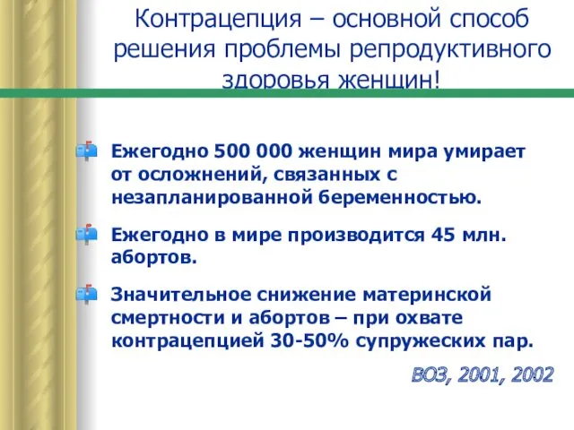 Контрацепция – основной способ решения проблемы репродуктивного здоровья женщин
 Перитонит. Классификация
 Анатомо-физиологические особенности, методика исследования органов дыхания у детей. Методы обследования
 Сердечно-легочная реанимация. Терминальные состояния
 Болезни и травмы органов дыхания
 Корнеальный синдром. Общие симптомы кератитов. Герпетические кератиты. Орбитальные и бульбарные боли. Иридоциклит
 Бронхообструктивті синдром Желшешек Қызылша қызамық
 Эндодонтическая подготовка к проведению хирургических методов лечения заболеваний пульпы и периодонта
 Задачи и особенности восстановительной хирургии челюстно-лицевой области. Дефекты и деформации. Лекция № 8
 Пластмассы (полимеры)
 Патогенез лейкозов
 Жылдам дамитын пародонт қабынуы
 Всемирный день здоровья
 Жүрек ақаулары
 Составление схем диспансерного наблюдения у курируемых хронических больных
 Гнойная хирургия конечностей
 Перинатальная охрана плода и новорожденного
 Кишечная форма муковисцидоза у детей
 Depression and the consumption of alcohol and tobacco among youth, cross-sectional analysis
 Актуальные вопросы ВИЧ/СПИД
 Жыныстық гормондар және жыныстық тәртіп
 Жаңа туылған нәрестенің геморрагиялық синдром