- Leptospirosis

Содержание
- 2. ETIOLOGY: The infectious agent – leptospira ( F. Spirochaetacea K. Leptospira) also is subdivided on pathogenic
- 5. Optimal conditions of the growth on the medium with addition 5 - 10 % serum of
- 6. L. have 2 antigenes: - genuspecific (deep) - and typespecific (surface). During illness will be derivated
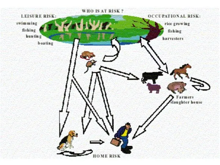
- 8. DWELLING-PLACE OF wild small rodents mice, rats hedgehogs ect.)
- 10. Seasonal rise - summur- autumn ( superactivity rodents, frequent contacts of the people to FRESH water)
- 11. The main pathogene of an icteric leptospirosis - L. icterohaemorragie ( but the icteric form maybe
- 12. 3. Reproduction in these bodies and secondary bacteriemia, which results in appearance of a SEPTIC phase
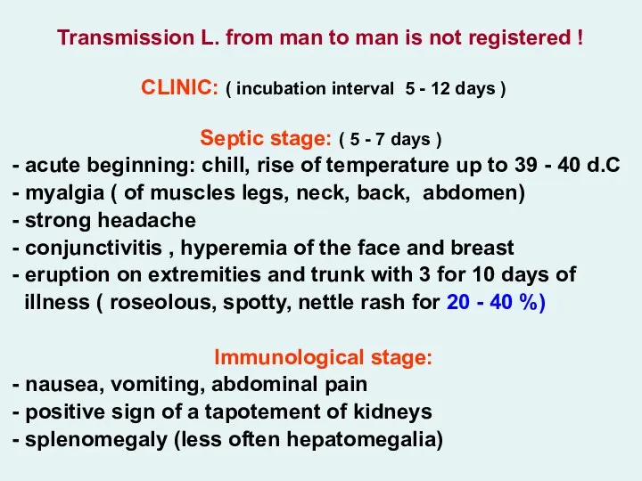
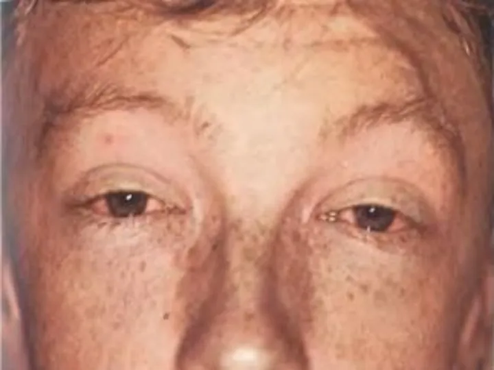
- 14. Transmission L. from man to man is not registered ! CLINIC: ( incubation interval 5 -
- 21. appearance meningeal of a syndrome with a pleocytosis and by increase of protein in CSF hyperleukocytosis
- 24. Leptospirosis can cause jaundice
- 27. - increase conjugated (direct) bilirubin at moderate rise АLТ (are not higher 2 - 4 norms)
- 28. LABORATORY DIAGNOSIS: - microscopy of a blood in a dark field (positive to 10 %) microscopy
- 29. - the icteric form: virus hepatitises, yellow fever, malaria, visceral leishmaniasis, yersiniosis, pseudotuberculosis, hemorrhagic fevers etc.
- 30. - at acute renal unsufficiency – diuretics (at once osmotic, at anuria - saluretics, but if
- 31. ANTHRAX – A. The acute infectious zoonotic disease described by development for the man of a
- 32. ETIOLOGY: Bacillus anthracis - large rod with equal edges 3-8 microns of length and 1-1.5 microns
- 36. Thermostable somatic polysaccharide the antigene is durably saved in corpses (is discovered by response of a
- 37. - eating corpses perished animals (predators) - through stings of insects (gadflies, horseflies, flies) Auxiliary source
- 38. Cases of man-to man transmission is not registered ! More often are sick cattle-breeder. The case
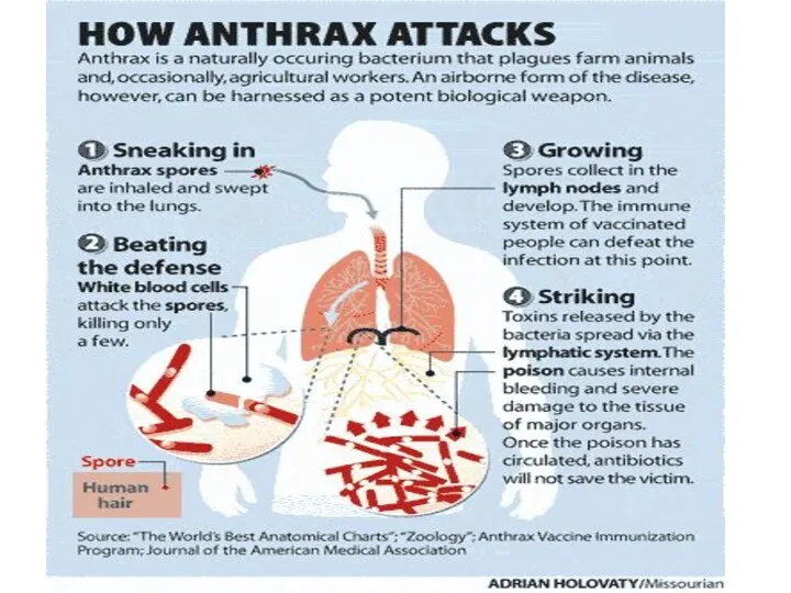
- 39. 3. The primary anthracic pneumonia does not develop! At aerogenic way of infection of spores will
- 40. CLINIC: (incubation interval from 2 about 14 days) The localized form of disease: in a place
- 52. Simultaneously around of a ulcer the edema considerably exceeding size of a ulcer is shaped. At
- 55. The generalized form А. - The incubation interval can be reduced about 1 day Acute beginning
- 56. Hemorrhagic meningitis resulting from inhalation anthrax.
- 57. Anthrax: blood clot passed from anus http://pathmicro.med.sc.edu/ghaffar/zoonoses.htm
- 58. Chest radiograph showing widened mediastinum resulting from inhalation anthrax.
- 59. Inhalational anthrax initially causes influenza-like symptoms, which progress to high fevers and severe respiratory distress. Typical
- 60. Inhalational anthrax - Mediastinal widening
- 61. - the patients durably save consciousness in despite of gravity of a state, except for cases
- 62. LABORATORY DIAGNOSIS - contents of pustules, discharge of an ulcer, blood, urine, sputum, stool, vomitive masses,
- 63. - tetracyclin 8 - 10 mg/kg q6h РО doxycyclin 2 mg/kg q12h РО chloramphenicol 8-10 mg/kg
- 64. PROPHYLAXIS: Obligatory hospitalization of the patients (EHI) Overseeing contacts within 14 days Emergency chemoprophilaxis contacts at
- 69. Скачать презентацию
ETIOLOGY:
The infectious agent – leptospira ( F. Spirochaetacea K. Leptospira) also
The infectious agent – leptospira ( F. Spirochaetacea K. Leptospira) also


Optimal conditions of the growth on the medium with addition
Optimal conditions of the growth on the medium with addition
L. have 2 antigenes: - genuspecific (deep)
- and typespecific
- and typespecific
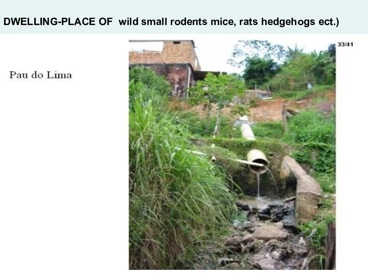
DWELLING-PLACE OF wild small rodents mice, rats hedgehogs ect.)
DWELLING-PLACE OF wild small rodents mice, rats hedgehogs ect.)

Seasonal rise - summur- autumn ( superactivity rodents, frequent contacts of
The main pathogene of an icteric leptospirosis
- L. icterohaemorragie (
- L. icterohaemorragie (
3. Reproduction in these bodies and secondary bacteriemia, which results in

Transmission L. from man to man is not registered !
CLINIC: (
CLINIC: (


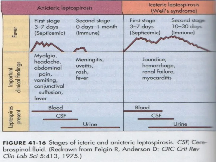

appearance meningeal of a syndrome with a pleocytosis
and by
and by


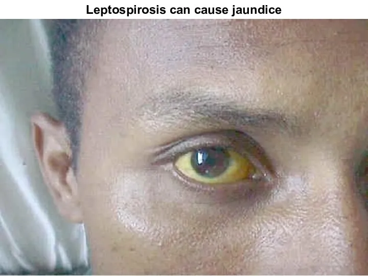
Leptospirosis can cause jaundice
Leptospirosis can cause jaundice


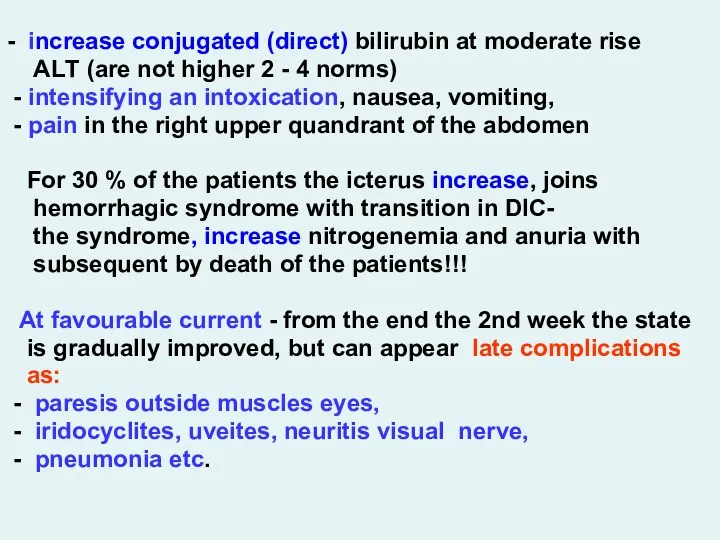
- increase conjugated (direct) bilirubin at moderate rise
АLТ
- increase conjugated (direct) bilirubin at moderate rise
АLТ
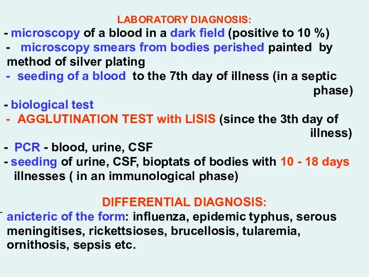
LABORATORY DIAGNOSIS:
- microscopy of a blood in a dark field (positive
- microscopy of a blood in a dark field (positive
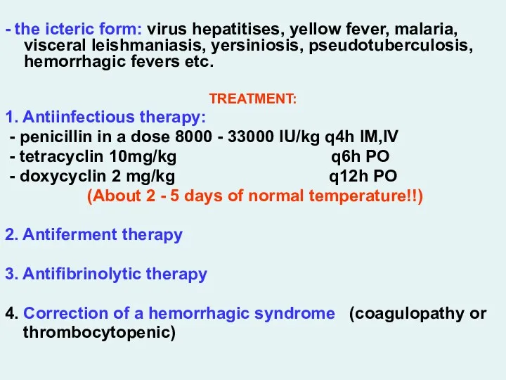
- the icteric form: virus hepatitises, yellow fever, malaria, visceral
- the icteric form: virus hepatitises, yellow fever, malaria, visceral
- at acute renal unsufficiency – diuretics (at once osmotic,
- at acute renal unsufficiency – diuretics (at once osmotic,
ANTHRAX – A.
The acute infectious zoonotic disease described
ANTHRAX – A.
The acute infectious zoonotic disease described
ETIOLOGY:
Bacillus anthracis - large rod with equal edges 3-8
Bacillus anthracis - large rod with equal edges 3-8


Thermostable somatic polysaccharide the antigene is durably saved in corpses
Thermostable somatic polysaccharide the antigene is durably saved in corpses
- eating corpses perished animals (predators)
- through stings of insects (gadflies,
- through stings of insects (gadflies,
Cases of man-to man transmission is not registered !
Cases of man-to man transmission is not registered !
3. The primary anthracic pneumonia does not develop!
At aerogenic way
At aerogenic way
CLINIC: (incubation interval from 2 about 14 days)
The localized form
CLINIC: (incubation interval from 2 about 14 days)
The localized form


Simultaneously around of a ulcer the edema considerably exceeding size
Simultaneously around of a ulcer the edema considerably exceeding size


The generalized form А.
- The incubation interval can be reduced
- The incubation interval can be reduced
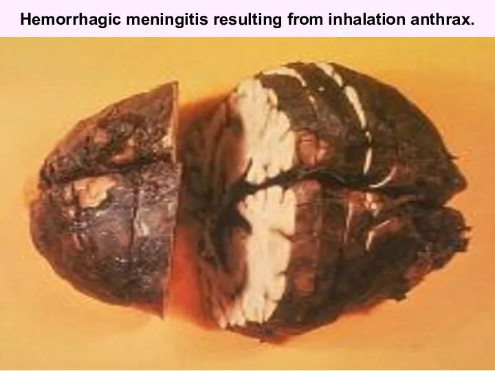
Hemorrhagic meningitis resulting from inhalation anthrax.
Hemorrhagic meningitis resulting from inhalation anthrax.
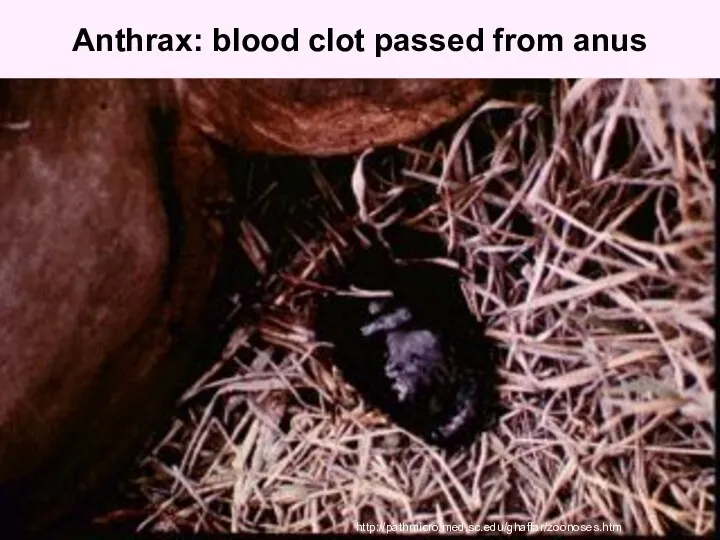
Anthrax: blood clot passed from anus
http://pathmicro.med.sc.edu/ghaffar/zoonoses.htm
Anthrax: blood clot passed from anus
http://pathmicro.med.sc.edu/ghaffar/zoonoses.htm
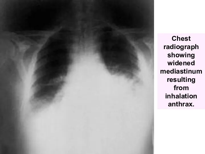
Chest radiograph showing widened mediastinum resulting from inhalation anthrax.
Chest radiograph showing widened mediastinum resulting from inhalation anthrax.
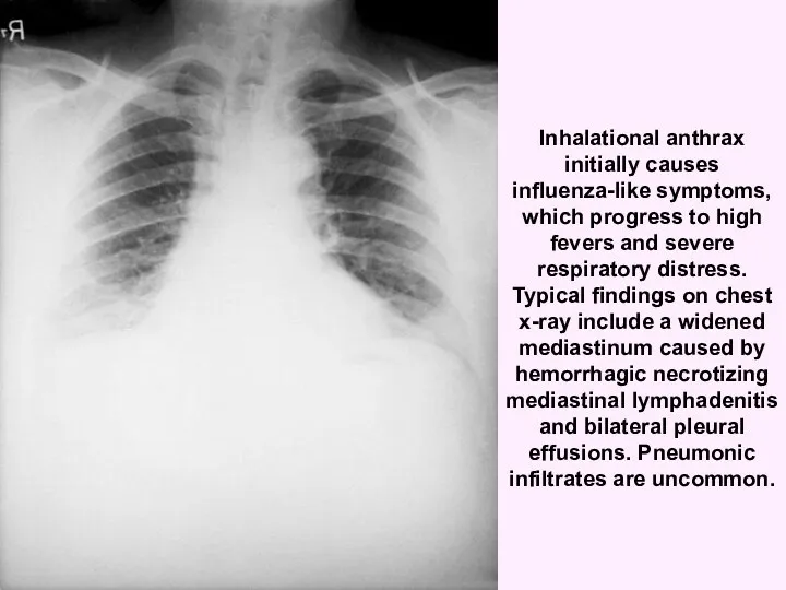
Inhalational anthrax initially causes influenza-like symptoms, which progress to high fevers
Inhalational anthrax initially causes influenza-like symptoms, which progress to high fevers
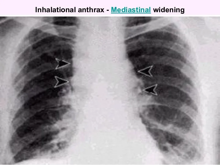
Inhalational anthrax - Mediastinal widening
Inhalational anthrax - Mediastinal widening
- the patients durably save consciousness in despite of
gravity
gravity
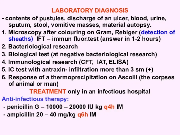
LABORATORY DIAGNOSIS
- contents of pustules, discharge of an ulcer, blood,
- contents of pustules, discharge of an ulcer, blood,
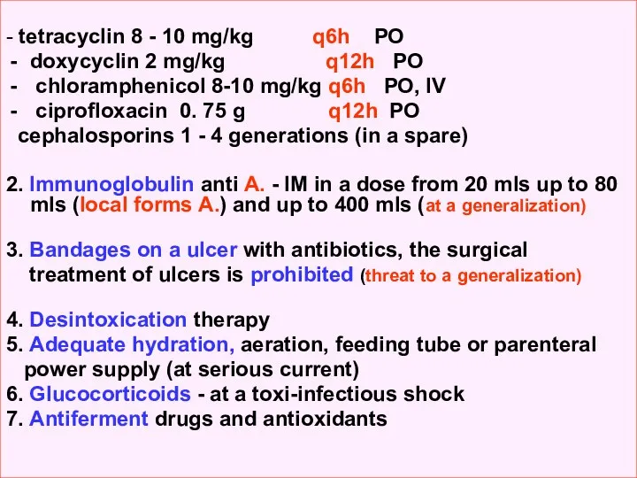
- tetracyclin 8 - 10 mg/kg q6h РО
doxycyclin 2 mg/kg
doxycyclin 2 mg/kg
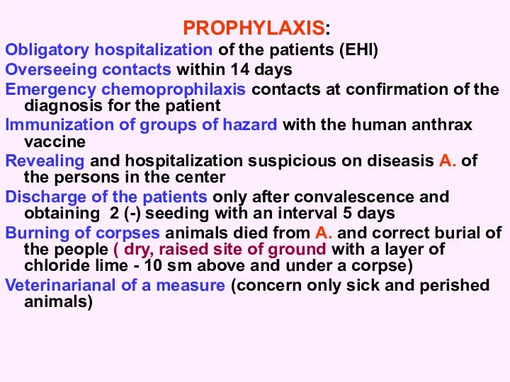
PROPHYLAXIS:
Obligatory hospitalization of the patients (EHI)
Overseeing contacts within 14 days
Emergency chemoprophilaxis
Obligatory hospitalization of the patients (EHI)
Overseeing contacts within 14 days
Emergency chemoprophilaxis


 Алкогольные и неалкогольные стеатогепатиты
 Диабеттік нефропатия диагнозын анықтау, емдеу қантты диабеттің кеш асқынулары
 Симптоматология ревматоидного артрита, остеоартроза, диффузных болезней соединительной ткани. (Тема 33)
 Современная стоматологическая клиника социальной направленности (на примере р.п. Кантемировка)
 Внутричерепная гипертензия
 Медленные инфекции. Механизм патогенеза
 Геморрагическая лихорадка с почечным синдромом
 Лапароскопия
 Клинические случаи в практике врача стоматолога
 История анестезиологии
 Развитие сестринского дела в России
 Дифференциальная диагностика заболеваний с респираторным синдромом
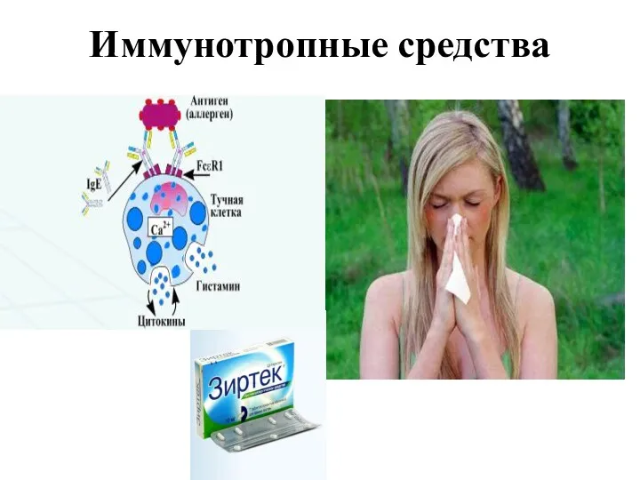 Иммунотропные средства
 Аллергия: диагностика, профилактика, лечение
 Балалар мекемелеріндегі санитарлық-техникалық жабдықтарға қойылатын гигиеналық талаптар. (Дәріс 19)
 12 жұп бас ми нервтері
 Болезни конъюнктивы
 Фармакоэкономика. Оценка экономической эффективности лекарственных средств
 Вырусные поражения половых органов
 Microscopic colitis
 Жасөспірім шақтағы ерте жүктіліктің алдын-алу
 Лицевой нерв
 Синдром длительного сдавления
 Қызметтік жүйелер туралы Анохиннің ілімі
 Бет аймағының ақауларын протездеу. Эктопротездер
 Расстройства воли и влечений, двигательные нарушения
 Методы исследования ВНД. Электроэнцефалография. Лекция 2
 Роль труда, спорта, социальных и биологических факторов на строение костей