- Микрофлора полости рта

Содержание
- 2. Основные группы бактерий вегетирующих в полости рта.
- 4. Типы дыхания бактерий Анаэробное Аэробное Факультативно-анаэробное дыхание
- 5. Gram Positive Gram Negative
- 6. The Gram negative cell envelope Covalently linked to the thin peptidoglycan is the Braun lipoprotein which
- 7. Gram Positive Cell Envelope (Figure 2) Covalently bound to the thick peptidoglycan are teichoic acid (their
- 8. 1. Эмаль и цемент зуба Кислотообразующие микробы: 1. Streptococcus mutans 2. Actinomyces spp. 3. Lactobacterium spp.
- 9. Электронная микроскопия стрептококков
- 10. Стрептококки
- 11. Гемолиз на кровяном агаре.
- 12. Бета-гемолитические колонии S.milleri на кровяном агаре
- 13. 2. Зубо-десневой желобок Грам-отрицательные бактерии. 1. Prevotella melaninogenica 2. Fusobacterium spp. Грам-положительные бактерии. 1.Peptostreptococcus spp. 2.Actinomyces
- 14. Превотеллы на кровяном агаре
- 15. Prevotella bivia
- 16. Мазок из чистой культуры бактероидов
- 17. Препарат из чистой культуры Fusobacteriun nucleatum
- 18. Фузобактерии и спирохеты.
- 19. Фузобактерии (окраска по Граму)
- 20. Фузобактерии на кровяном агаре
- 21. Актиномицеты
- 22. Рост чистой культуры Actinomyces israeli на кровяном агаре.
- 23. Пептострептококки
- 24. Пептострептококки в мазке из гнойного экссудата
- 25. 3. Спинка языка 1.Streptococcus salivarius 2. Streptococcus sanguis 3. Lactobacillus spp. 4. Leptotrichia buccalis 5. Candida
- 26. Морфология стрептококков
- 27. 4. Лакуны слизистой оболочки полости рта Грам-положительные бактерии. 1. Streptococcus sangus 2. Streptococcus mitis 3. Peptostreptococcus
- 28. Пептострептококки
- 29. Нокардии
- 30. Спирохеты
- 31. Спирохеты. Электронная микроскопия.
- 32. 5. Слизистая оболочка полости рта Грам-положительные бактерии 1. S.sanguis 2. S. mitis 3. Corynebacterium spp. Грам-отрицательные
- 33. Пропионибактерии
- 34. Коринебактерии
- 35. Дрожжеподобные грибы кандида.
- 36. Candida albicans (окраска по Граму)
- 37. Рост на питательной среде разных видов грибов кандида
- 38. Изолированные колонии Candida albicans и Candida tropicalis на плотной питательной среде.
- 39. Пути распространения одонтогенной инфекции
- 40. Диагностика одонтогенных инфекций
- 43. Скачать презентацию
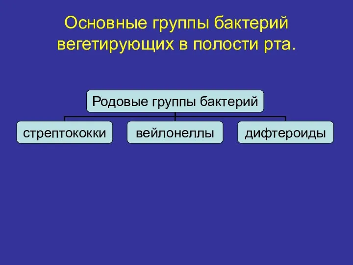
Основные группы бактерий вегетирующих в полости рта.
Основные группы бактерий вегетирующих в полости рта.
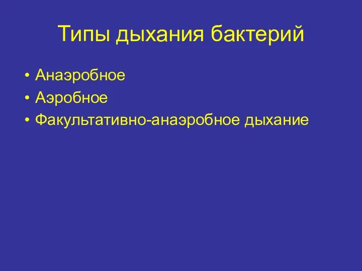
Типы дыхания бактерий
Анаэробное
Аэробное
Факультативно-анаэробное дыхание
Типы дыхания бактерий
Анаэробное
Аэробное
Факультативно-анаэробное дыхание
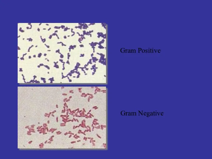
Gram Positive
Gram Negative
Gram Positive
Gram Negative
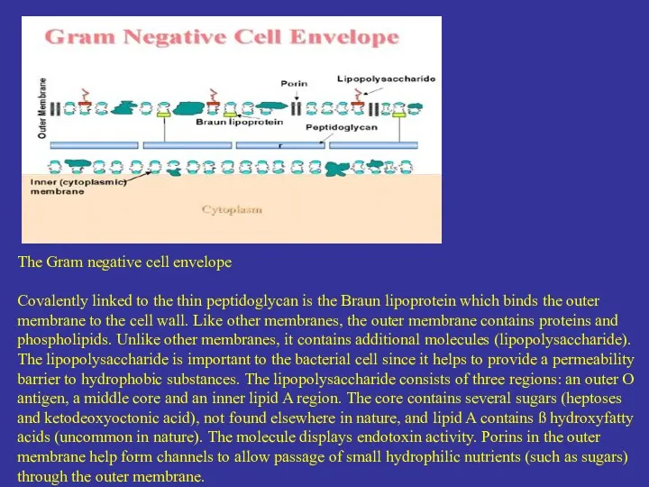
The Gram negative cell envelope
Covalently linked to the thin peptidoglycan
The Gram negative cell envelope
Covalently linked to the thin peptidoglycan
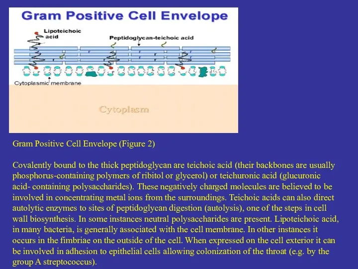
Gram Positive Cell Envelope (Figure 2)
Covalently bound to the thick peptidoglycan
Gram Positive Cell Envelope (Figure 2)
Covalently bound to the thick peptidoglycan
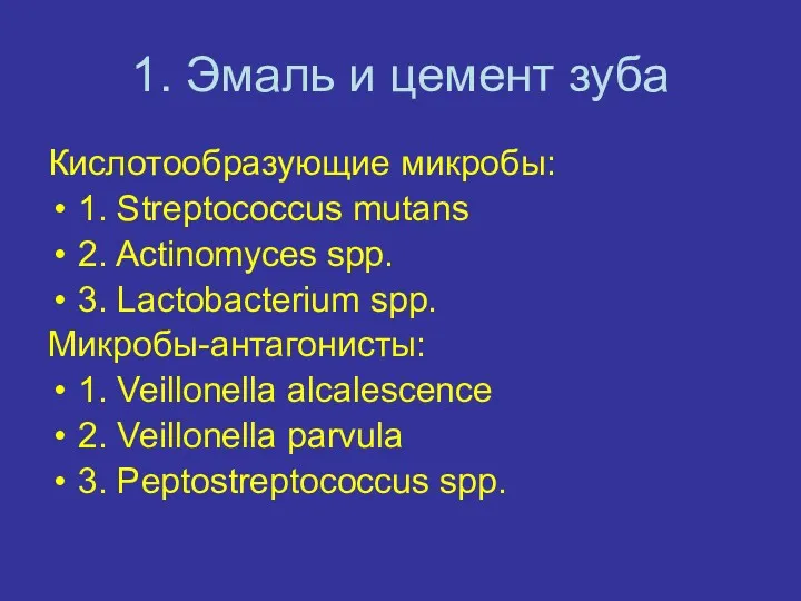
1. Эмаль и цемент зуба
Кислотообразующие микробы:
1. Streptococcus mutans
2. Actinomyces spp.
3. Lactobacterium
1. Эмаль и цемент зуба
Кислотообразующие микробы:
1. Streptococcus mutans
2. Actinomyces spp.
3. Lactobacterium
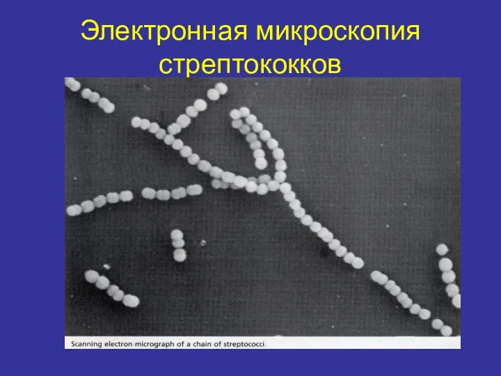
Электронная микроскопия стрептококков
Электронная микроскопия стрептококков
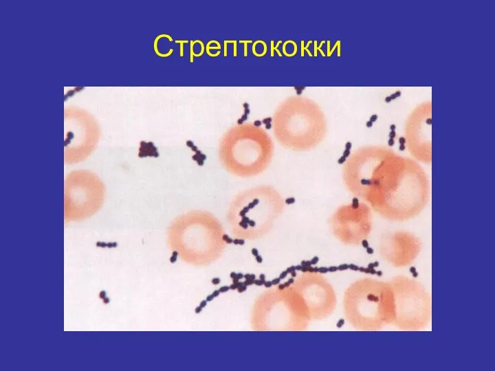
Стрептококки
Стрептококки
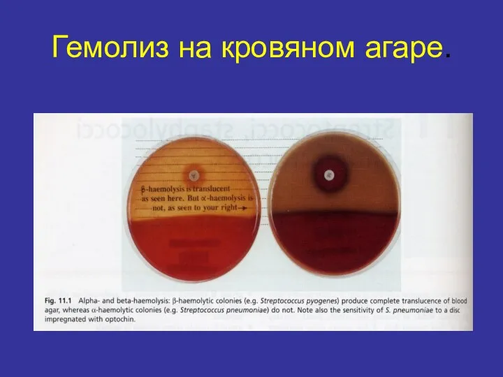
Гемолиз на кровяном агаре.
Гемолиз на кровяном агаре.
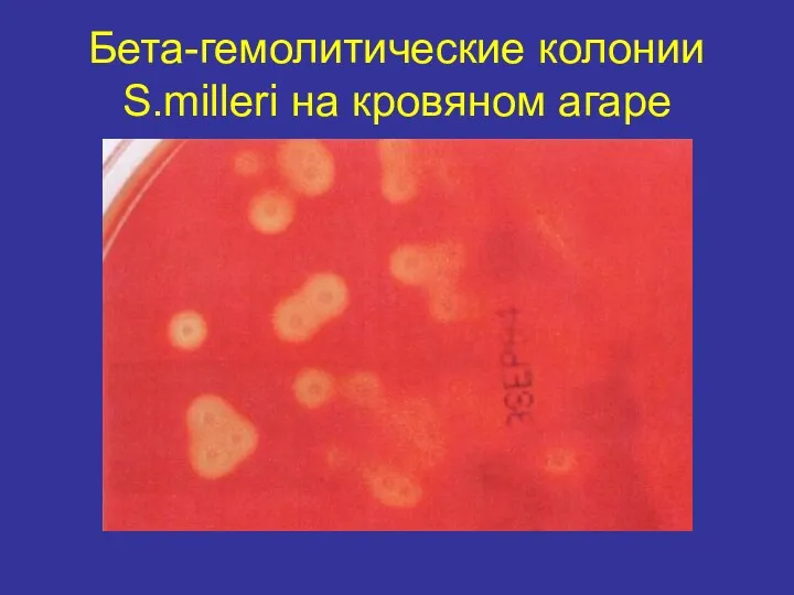
Бета-гемолитические колонии S.milleri на кровяном агаре
Бета-гемолитические колонии S.milleri на кровяном агаре
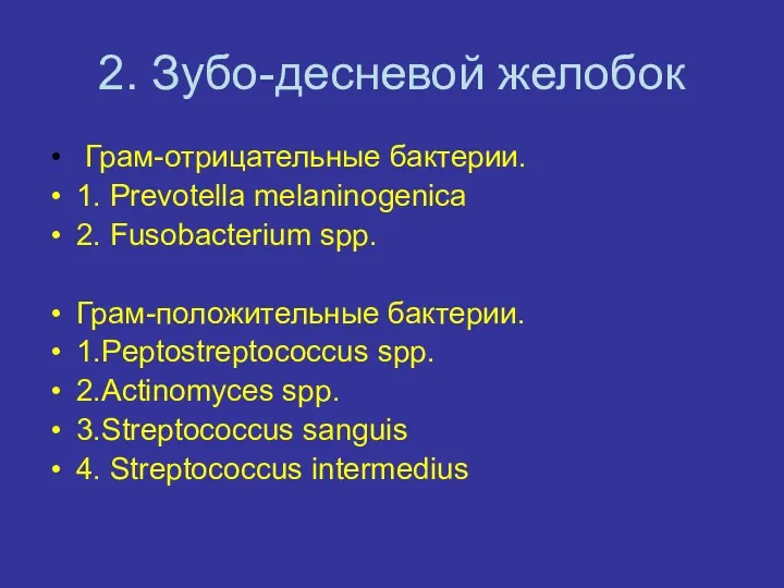
2. Зубо-десневой желобок
Грам-отрицательные бактерии.
1. Prevotella melaninogenica
2. Fusobacterium spp.
Грам-положительные бактерии.
1.Peptostreptococcus spp.
2.Actinomyces
2. Зубо-десневой желобок
Грам-отрицательные бактерии.
1. Prevotella melaninogenica
2. Fusobacterium spp.
Грам-положительные бактерии.
1.Peptostreptococcus spp.
2.Actinomyces
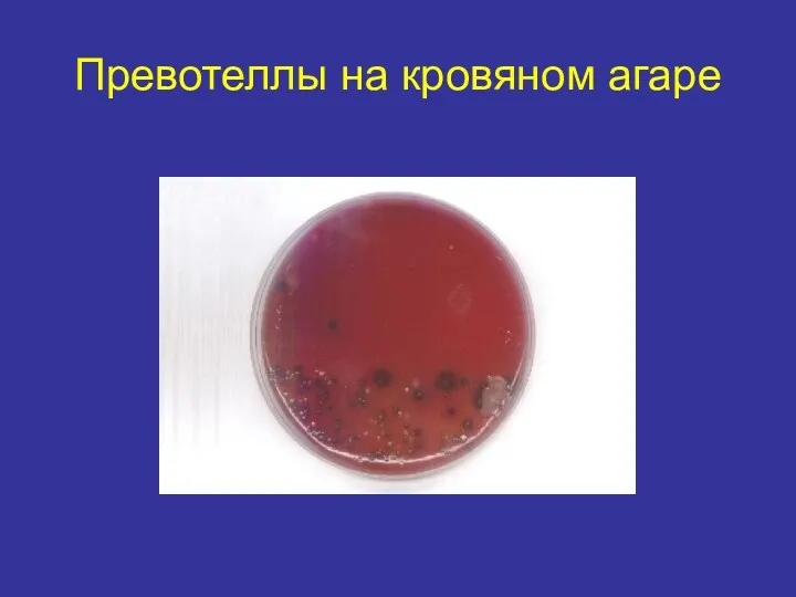
Превотеллы на кровяном агаре
Превотеллы на кровяном агаре
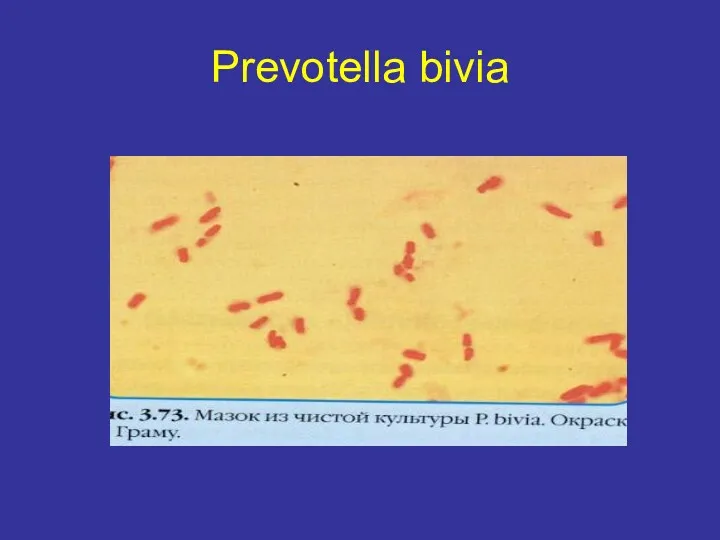
Prevotella bivia
Prevotella bivia
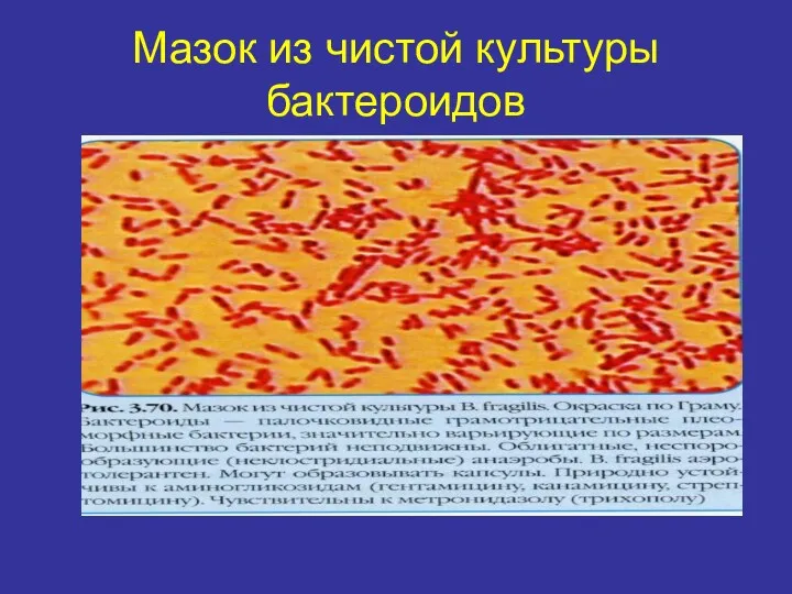
Мазок из чистой культуры бактероидов
Мазок из чистой культуры бактероидов
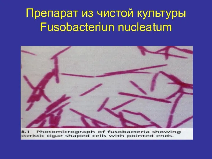
Препарат из чистой культуры Fusobacteriun nucleatum
Препарат из чистой культуры Fusobacteriun nucleatum
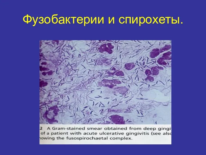
Фузобактерии и спирохеты.
Фузобактерии и спирохеты.
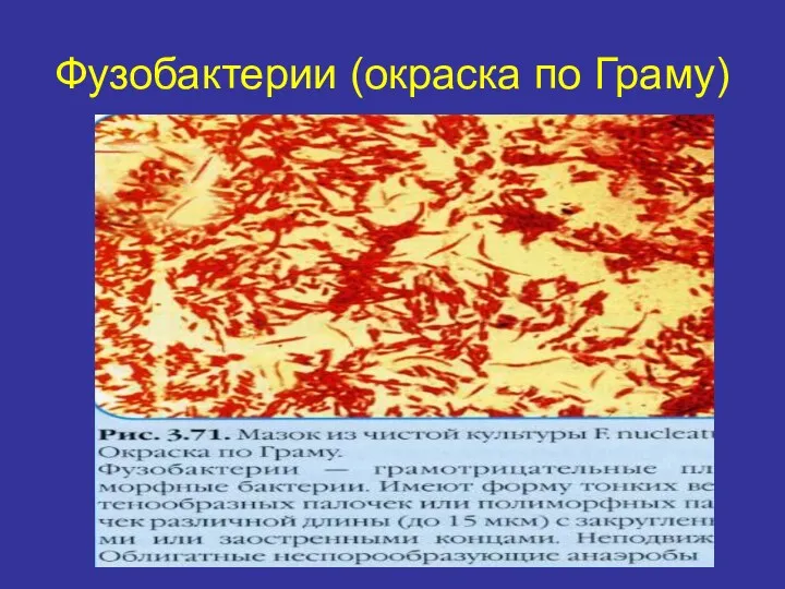
Фузобактерии (окраска по Граму)
Фузобактерии (окраска по Граму)

Фузобактерии на кровяном агаре
Фузобактерии на кровяном агаре
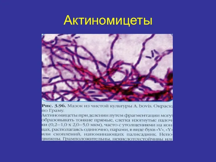
Актиномицеты
Актиномицеты
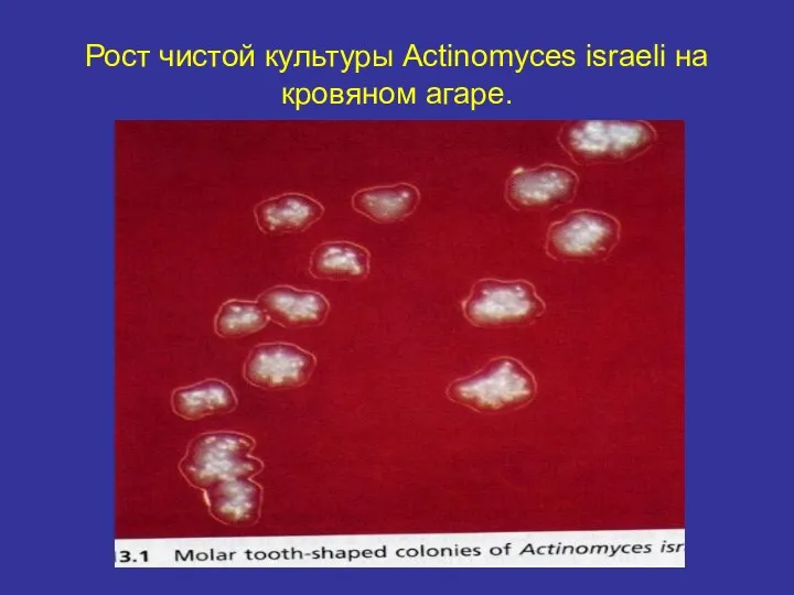
Рост чистой культуры Actinomyces israeli на кровяном агаре.
Рост чистой культуры Actinomyces israeli на кровяном агаре.
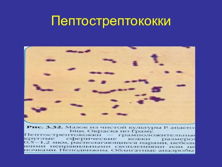
Пептострептококки
Пептострептококки
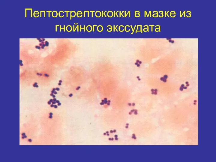
Пептострептококки в мазке из гнойного экссудата
Пептострептококки в мазке из гнойного экссудата
3. Спинка языка
1.Streptococcus salivarius
2. Streptococcus sanguis
3. Lactobacillus spp.
4. Leptotrichia buccalis
5. Candida
3. Спинка языка
1.Streptococcus salivarius
2. Streptococcus sanguis
3. Lactobacillus spp.
4. Leptotrichia buccalis
5. Candida
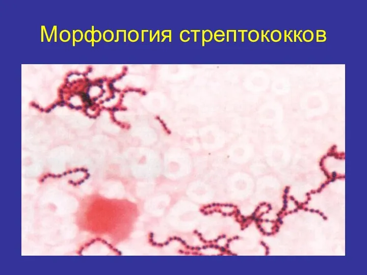
Морфология стрептококков
Морфология стрептококков
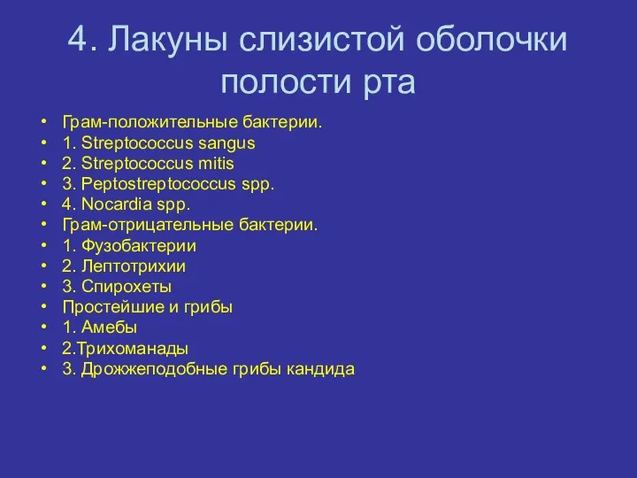
4. Лакуны слизистой оболочки полости рта
Грам-положительные бактерии.
1. Streptococcus sangus
2. Streptococcus mitis
3.
4. Лакуны слизистой оболочки полости рта
Грам-положительные бактерии.
1. Streptococcus sangus
2. Streptococcus mitis
3.
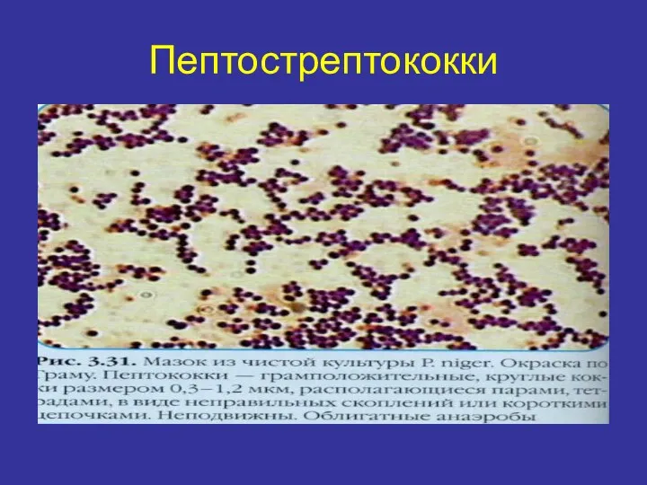
Пептострептококки
Пептострептококки
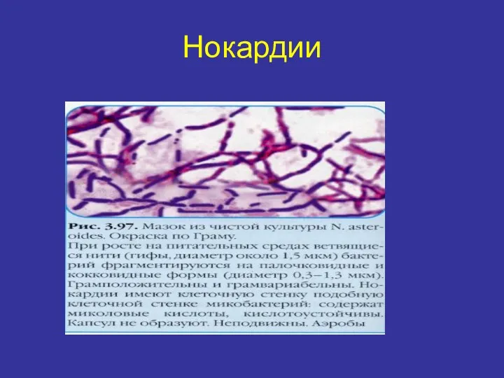
Нокардии
Нокардии
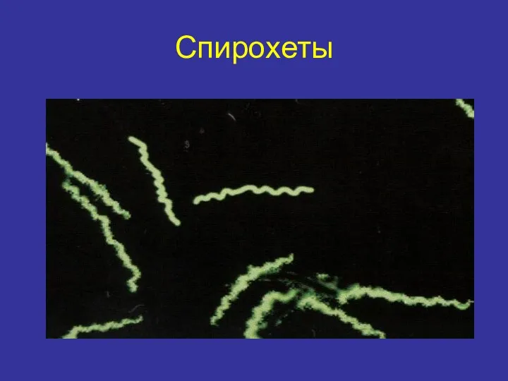
Спирохеты
Спирохеты
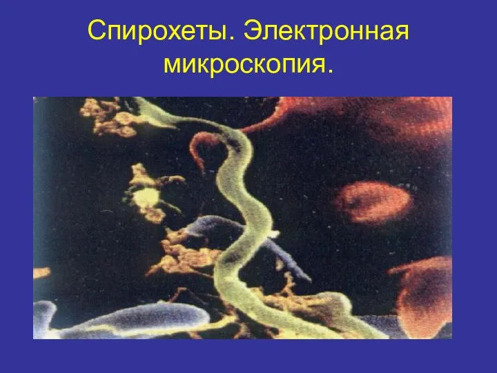
Спирохеты. Электронная микроскопия.
Спирохеты. Электронная микроскопия.
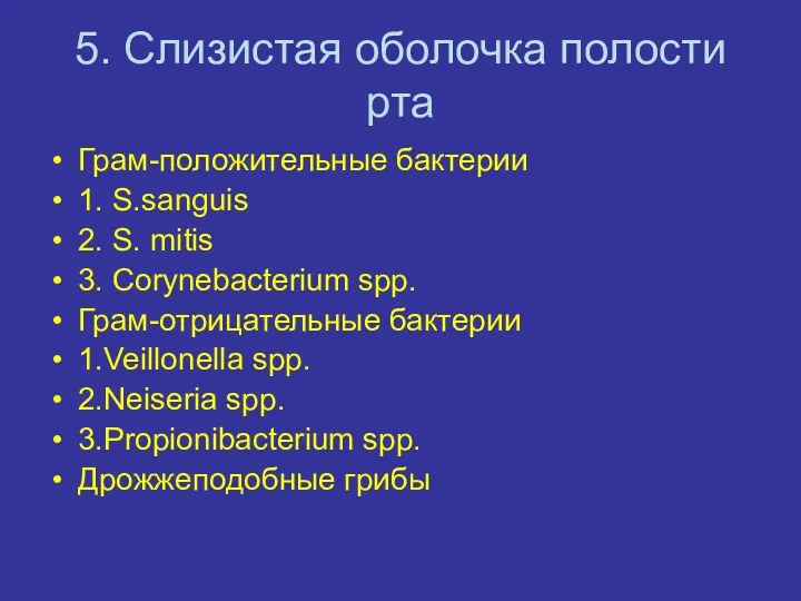
5. Слизистая оболочка полости рта
Грам-положительные бактерии
1. S.sanguis
2. S. mitis
3. Corynebacterium spp.
Грам-отрицательные
5. Слизистая оболочка полости рта
Грам-положительные бактерии
1. S.sanguis
2. S. mitis
3. Corynebacterium spp.
Грам-отрицательные
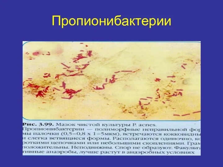
Пропионибактерии
Пропионибактерии
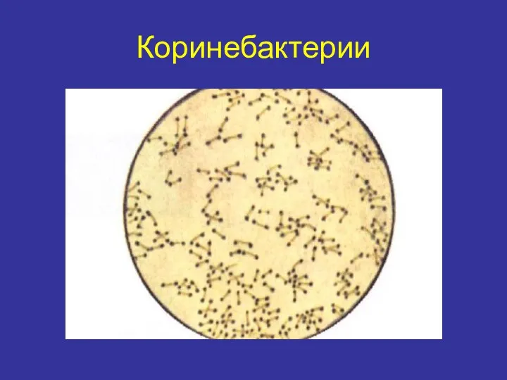
Коринебактерии
Коринебактерии
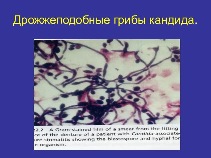
Дрожжеподобные грибы кандида.
Дрожжеподобные грибы кандида.
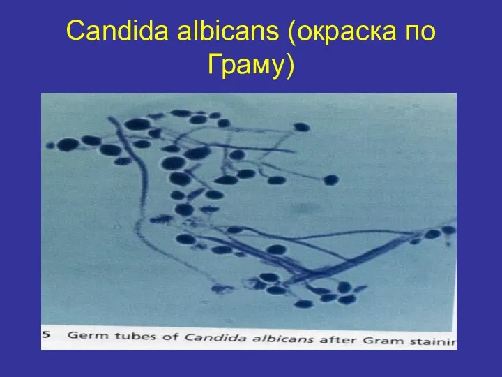
Candida albicans (окраска по Граму)
Candida albicans (окраска по Граму)
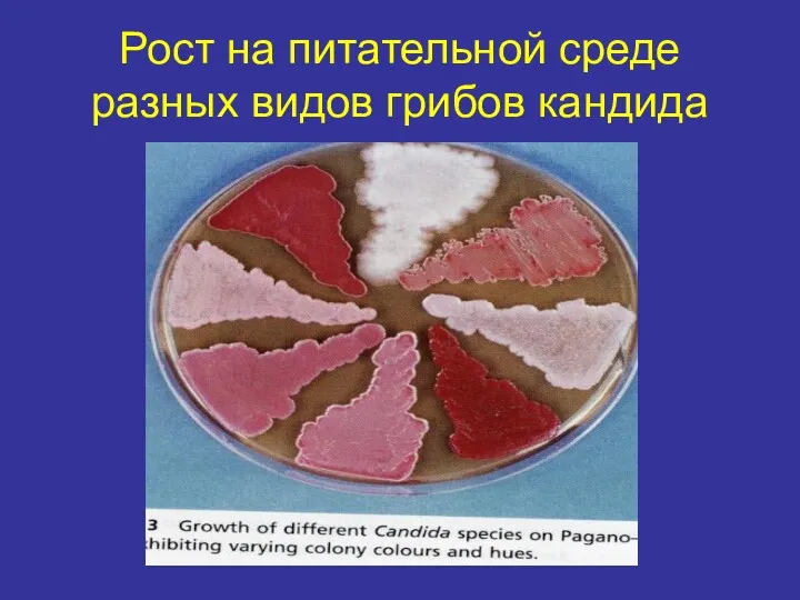
Рост на питательной среде разных видов грибов кандида
Рост на питательной среде разных видов грибов кандида
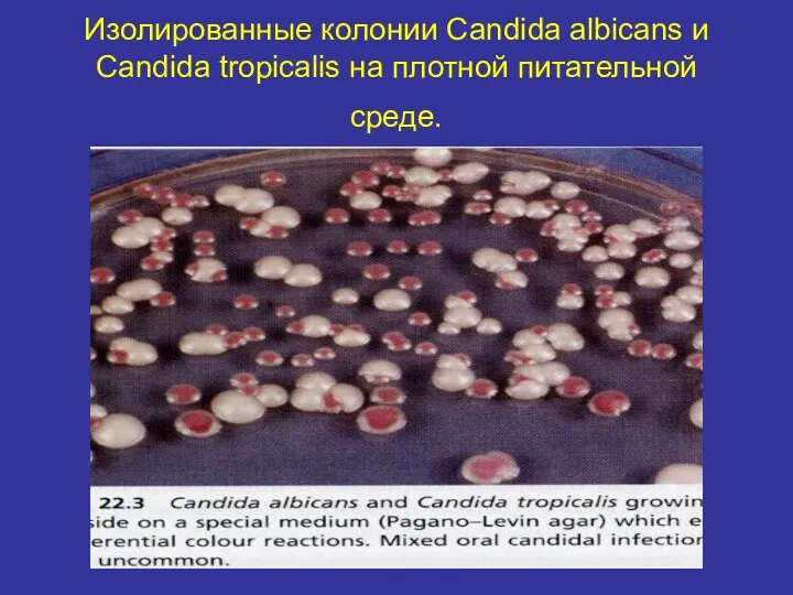
Изолированные колонии Candida albicans и Candida tropicalis на плотной питательной среде.
Изолированные колонии Candida albicans и Candida tropicalis на плотной питательной среде.
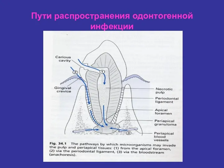
Пути распространения одонтогенной инфекции
Пути распространения одонтогенной инфекции
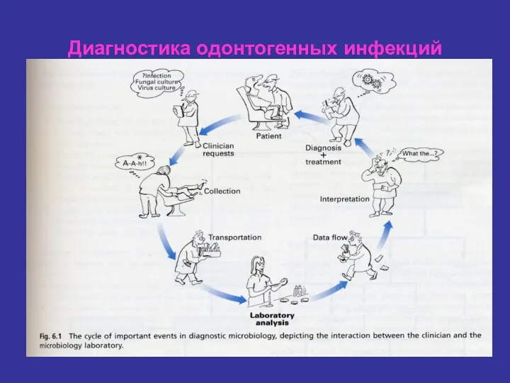
Диагностика одонтогенных инфекций
Диагностика одонтогенных инфекций

 Недонашивание и перенашивание беременности
 Химические ожоги пищевода у детей
 Черепно-мозговая травма
 Лекция ОП некроз
 Оценка эффективности программ профилактики и скрининга
 Принципы терапии инвазивных кандидозов в ОРИТ
 Исследование больных с патологией органов кроветворения
 Регуляция кровообращения
 Опухолеподобные заболевания костей. Дифференциальная диагностика
 Экзема дегеніміз - эпидермис пен дерманың қабынуы
 Неотложные состояния при сахарном диабете типа 1
 Болезнь Крона
 Система регуляции агрегатного состояния крови. Свертывание крови
 Гигиена труда медицинских работников
 Патронаж здорового новорожденного на дому с оформлением учебной амбулаторной карты
 Современные липосомальные противоопухолевые препараты
 Применение Конусно-лучевой компьютерной томографии(КЛКТ) в амбулаторной стоматологической практике
 Аварийные ситуации в стоматологии. Пути профилактики
 Внутриутробное развитие организма. Развитие после рождения
 Подагра. Подагритческий артрит
 Диагностика, лечение и профилактика бактериального вагиноза
 Диагностика и лечение талассемии
 Російські лікарі-філософи ХІХ століття
 Организация амбулаторно-поликлинической терапевтической службы в России
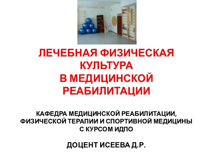 Лечебная физическая культура в медицинской реабилитации
 Транквилизаторы. Болеутоляющие средства. Седативные средства
 Анестезия в нейрохирургии
 Средства, влияющие на желудочно-кишечный тракт