- Radiospectroscopic research methods 4

Содержание
- 5. The proton-proton couplings in benzene are typically 7-9 Hz for Jortho, 2-3 Hz for Jmeta and
- 6. First Order Coupling Rules 1. Nuclei must be chemical shift nonequivalent to show obvious coupling to
- 7. 4. Chemical shifts are usually reported in δ (units: ppm) so that the numeric values will
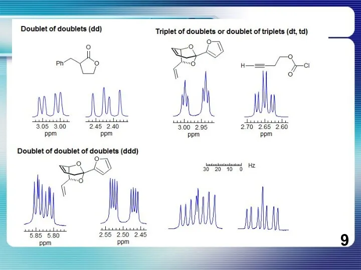
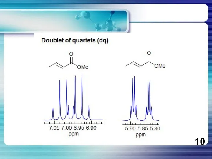
- 8. If all couplings are different, then the number of peaks is 2n for 1H, and the
- 11. Разрешение мультиплетов на приборах с разной рабочей частотой ))
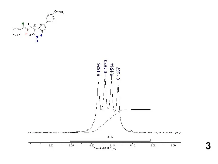
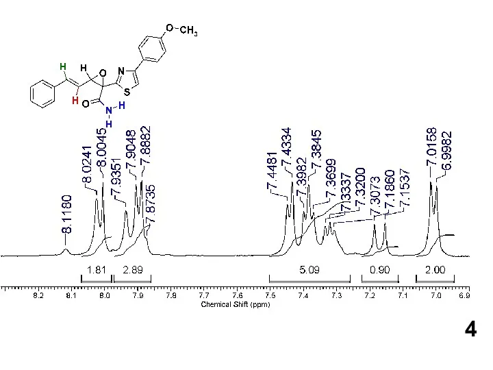
- 12. Protons or groups of protons form simple multiplets only if the chemical shift differences between the
- 13. АВ и АХ паттерны
- 15. The distinction between an AB q and a regular q is not always trivial. In fact,
- 17. Graphical method for determining the position of a leaning coupled partner. The point Q is the
- 18. Shown above is the 60 MHz spectrum of Abel's ketone in CDCl3 solution. There are three
- 19. Лирическое отступление об описании ЯМР спектров
- 20. How to report an AB quartet. Journals require that NMR spectra be reported in text format.
- 21. 13С ЯМР спектры
- 27. Скачать презентацию

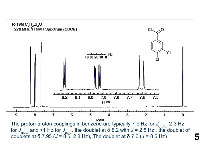
The proton-proton couplings in benzene are typically 7-9 Hz for Jortho, 2-3
The proton-proton couplings in benzene are typically 7-9 Hz for Jortho, 2-3
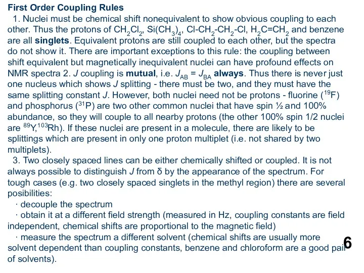
First Order Coupling Rules
1. Nuclei must be chemical shift nonequivalent to
First Order Coupling Rules
1. Nuclei must be chemical shift nonequivalent to
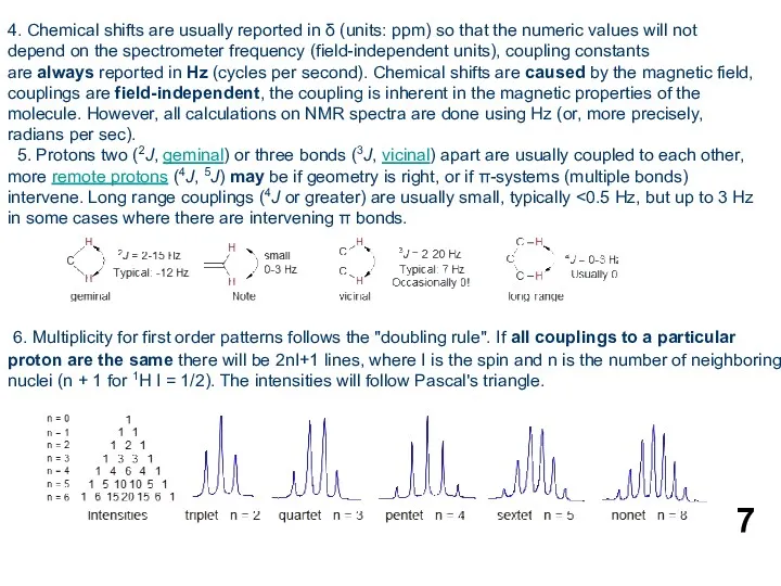
4. Chemical shifts are usually reported in δ (units: ppm) so
4. Chemical shifts are usually reported in δ (units: ppm) so
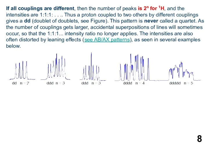
If all couplings are different, then the number of peaks is
If all couplings are different, then the number of peaks is
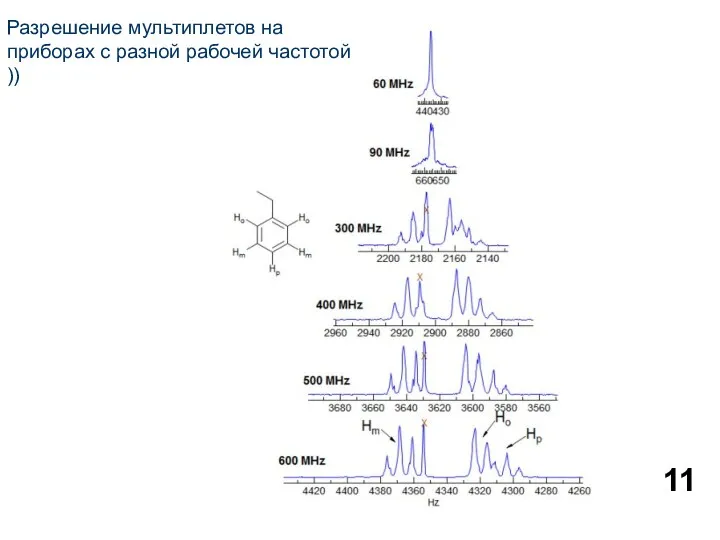
Разрешение мультиплетов на приборах с разной рабочей частотой ))
Разрешение мультиплетов на приборах с разной рабочей частотой ))
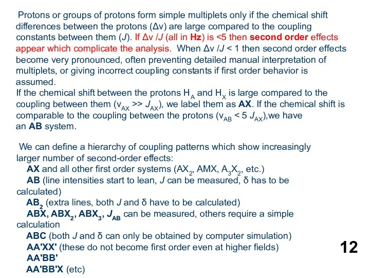
Protons or groups of protons form simple multiplets only if the
Protons or groups of protons form simple multiplets only if the
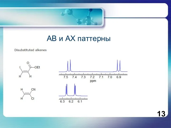
АВ и АХ паттерны
АВ и АХ паттерны
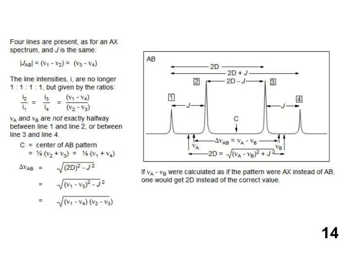
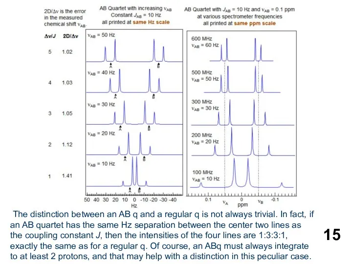
The distinction between an AB q and a regular q is
The distinction between an AB q and a regular q is
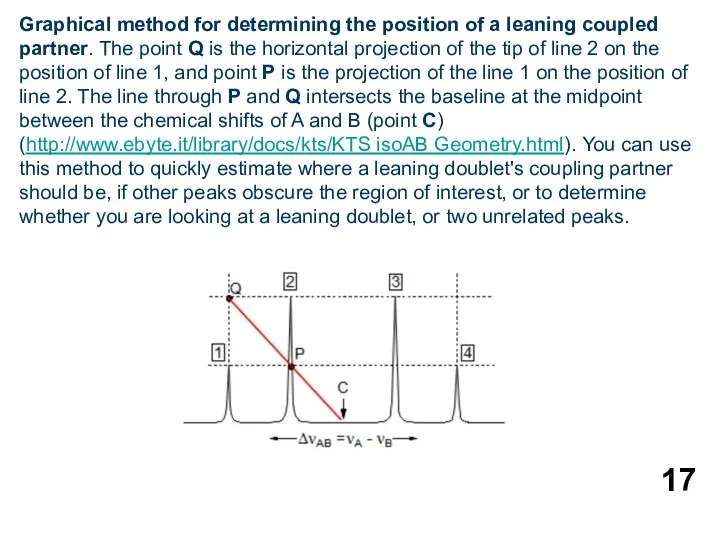
Graphical method for determining the position of a leaning coupled partner.
Graphical method for determining the position of a leaning coupled partner.
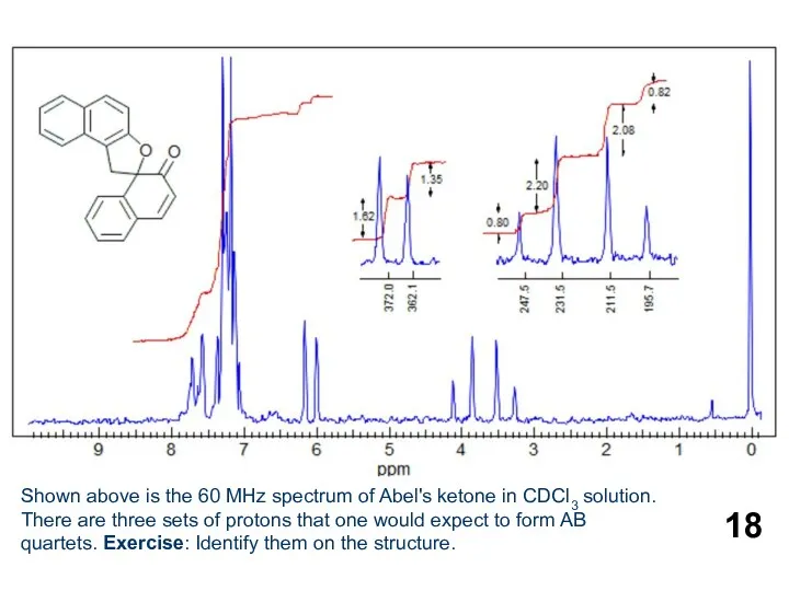
Shown above is the 60 MHz spectrum of Abel's ketone in
Shown above is the 60 MHz spectrum of Abel's ketone in
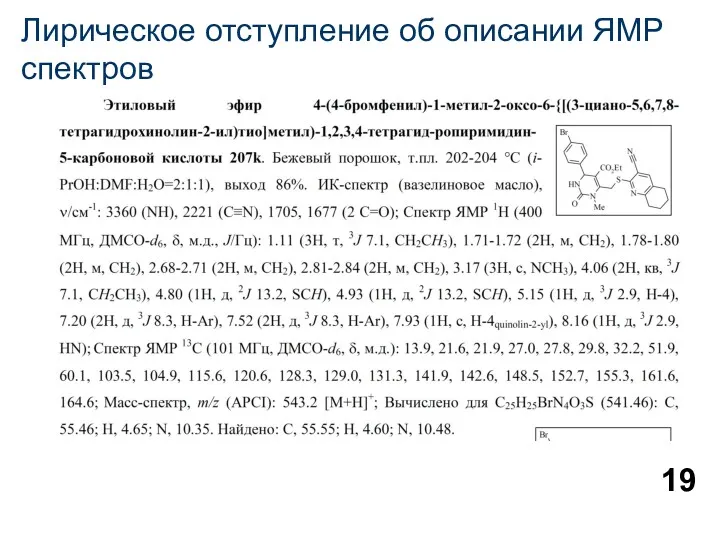
Лирическое отступление об описании ЯМР спектров
Лирическое отступление об описании ЯМР спектров
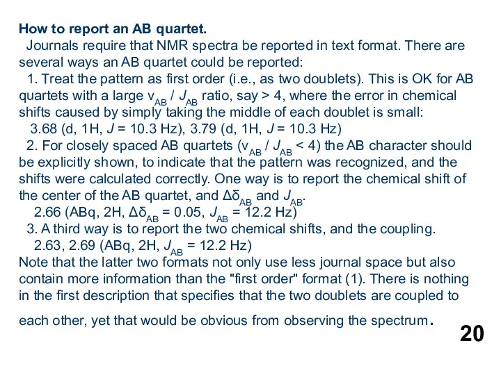
How to report an AB quartet.
Journals require that NMR spectra be
How to report an AB quartet.
Journals require that NMR spectra be
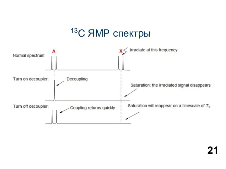
13С ЯМР спектры
13С ЯМР спектры


 Желтуха новорожденных
 Химиотерапия. Основные принципы химиотерапии
 Дети с задержкой психического развития (ЗПР)
 Сказ (rabies)
 Костномозговое кроветворение. Нормы крови. Схема кроветворения
 Этапы определения потребности в медицинском имуществе
 Алкоголизм и его влияние на развитие здоровой личности
 Побочные эффекты лекарственных вещевств
 УЗИ при беременности
 Общие сведения об алалии
 Воспалительные заболевания кишечника
 Лечение деменции
 Биологическое действие радиации
 Изменения состояния организма студентов под влиянием различных режимов и условий обучения
 Хранение и транспортирование товаров медицинского назначения на всех этапах товародвижения
 Проблема подросткового алкоголизма
 Неэффективность антимикробной терапии в хирургическом стационаре. Биопленочные инфекции
 Анафилактический шок. Неотложная помощь. Интенсивная терапия
 Рак слизистой оболочки полости рта. Клиника, диагностика, лечение
 Гигиеническое обеспечение занятий физической культурой и спортом. Утомление
 Бережливая поликлиника
 Введение в изучение нервной системы. Функциональная анатомия спинного и головного мозга
 Аллергические конъюнктивиты: классификация, клиника, лечение
 Антибіотикопрофілактика у хірургії
 Протездерді өңдеу технологиясы. Абразивті материалдар
 Патологическая стираемость генерализованного типа
 Көпұрықпен және көпбосанушы әйелдерге босанудан кейінгі қан кетудің алдын алу үшін Пабал және окситоцин препаратын қолдану
 Менингиты. Клиническая картина всех менингитов