Слайд 3BOTULISM
Ptosis in upper eyelids
Слайд 4HYPOTONIA
Decrease in muscle tone of infant
Слайд 5BOTULISM
Complete muscle paralysis in body of ducks
Слайд 8ACTINOMYCOSIS
Cattle: Lumpy jaw
Слайд 9ACTINOMYCOSIS
Cattle: Lumpy jaw
Слайд 10ACTINOMYCOSIS
Human: Cervicofacial form
Слайд 18RABIES
A- Rabid cattle: Profuse salivation & hoarse bellow
Слайд 20RABIES
Infected man restrained in a hospital
Слайд 21RABIES
Bullet shaped virus by electron microscope
Слайд 24erythematous lesions with central depression
MILKER’S NODULE
Слайд 25areas of swelling and reddening on teat
Слайд 27MANGE
Acute lesion of mange in man
Слайд 28MANGE
Acute lesion of mange in man
Слайд 30MANGE
Mange in animals: Alopecia & wrinkled skin
Слайд 41ECHINOCOCCOSIS
Rodent: Healthy & infested liver
Слайд 43ECHINOCOCCOSIS
Hydaited cyst in lung
Слайд 47TRICHINELLOSIS
pink eye:edema in the upper eyelids and subconjunctival hemorrhage due to migration
of Trichinella spirals larva
Слайд 48TRICHINELLOSIS
Trichenella spirals larva in pork muscle by trichinoscope
Слайд 51DIPHYLLOBOTHRIASIS
Colonoscopy revealed a long, moving tapeworm, Diphyllobothrium latum, located in the intestine
Слайд 54FASCIOLIASIS
Liver showing adult liver fluke
Слайд 57HUMAN PLAGUE
Ulcerated flea bite caused by Yersinia pestis bacteria
Слайд 58HUMAN PLAGUE
Enlargement in cerphical L.N
Слайд 59HUMAN PLAGUE
SEPTECIMIC FORM :
Congestion S.C blood vessels and cutaneous petechiae
Слайд 63YELLOW FEVER
- Jaundice – Hepatitis in man
Слайд 67LEISHMANIASIS
Leishmania form of Leishmania spp
Слайд 68LEISHMANIASIS
Leptomonad form of Leishmania spp
Слайд 69
LEISHMANIASIS
Cutaneous form : clear cut raised lesion
Слайд 71LEISHMANIASIS
Visceral form :
Abdominal distention due to hepatospleenomegaly
Слайд 74Trypanosomiasis
Tse tse fly
Loss of weight
Edema of the eye
Слайд 76RAT BIT FEVER
Maculopapular rash affecting man fingers
Слайд 78RAT BIT FEVER
Maculopapular rash affecting man fingers
Слайд 80ORF
-Pus-like sores on a goat's mouth
Слайд 82BARTONELLOSIS
Unilateral lymphadenitis
Слайд 83BARTONELLOSIS
Unilateral lymphadenitis
Слайд 84BARTONELLOSIS
Unilateral lymphadenitis
Слайд 86HEPATIC CYSTECERCOSIS
Multiple cyst in the liver
Слайд 87CYSTECERCOSIS
the impact of Large cyst in the brain
Слайд 89SWINE ERYSIPELAS
Diamond skin in pig
Слайд 90SWINE ERYSIPELAS
Localized cutaneous form
Слайд 91SWINE ERYSIPELAS
Localized cutaneous form
Слайд 92SWINE ERYSIPELAS
Localized cutaneous form
Слайд 94SKIN TEST (HANGER ROSE TEST)
FOR DETECTION OF CAT SCRATCH FEVER
ETIOLOGIC AGENT:
GRAM
NEGATIVE BACILLI CALLED ROCHALIMAEA HENSELAE OR BARTONELLA HENSELAE.
Слайд 95Wood's lamp illumination
Fluorescence due to the pteridine secreted fungi
Used for detection of
ring worm
Causative agent : anthropophilic- geophilic- zoofilic
Слайд 961. Anthropophilic dermatophytes:
Fungi of human being and may be transmitted to animals
as:
Microsporum audouinii
Trichophyton rubrum
Trichophyton shoenleini
Trichophyton violaceum
Epidermophyton floccosum
2. Zoophilic dermatophytes:
Fungi of animals and may be transmitted to man as: Trichophyton verrucosum (primary of cattle)
Trichophyton equinum (primary of equines)
Microsporum canis (primary of dogs)
Trichophyton mentagrophytes (primary of rodents)
Слайд 98TRICHINOSCOPE
Definition: a compression apparatus to squash a small sample of pork muscle
and a projector to display the image of tissue on a screen (Trichinella spp.)
Use: The trichinoscope is used for the detection of trichina cysts in pork muscle.
Causative agent: Trichinella spiralis (encysted larvae)
Слайд 100BLOCKAGE PHENOMENA
Blockage is a term used to describe occlusion of the flea’s
gut with a mass consisting of plague bacterium embedded within a biofilm produced by the bacterium itself
This blockage occurs at the point where the flea’s foregut (throat and esophagus) meets its midgut (stomach) in a spine-filled structure called the proventriculus
Слайд 101Infected rat flea(Xenopsylla cheopis) transmit bubonic form of human plague.
Casative agent : Yersinia
pestis
Слайд 103BIOLOGICAL CONTROL OF MOSQUITOES GUMBUSIA FISH (
Слайд 104– AERIAL APPLICATIONS(BACTERIAL-CONTROL )AGENTS

 Жатыр мойны эрозиясы
 Криомассаж. Современная технология закаливания и оздоровления детей
 Дискинезии желчевыводящих путей у детей
 Казеозды пневмония
 Система гемостаза. Геморрагические синдромы
 Дифференциальная диагностика суставного синдрома
 Здоровье человека. Факторы, определяющие здоровье человека. Основные причины болезней. Оценка состояния здоровья человека
 Экстрагенитальная патология и беременность
 Лазеротерапия. Организация кабинета. Матричные лазеры. Механизмы НИЛИ. Методы и методики
 Деструктивный туберкулез легких
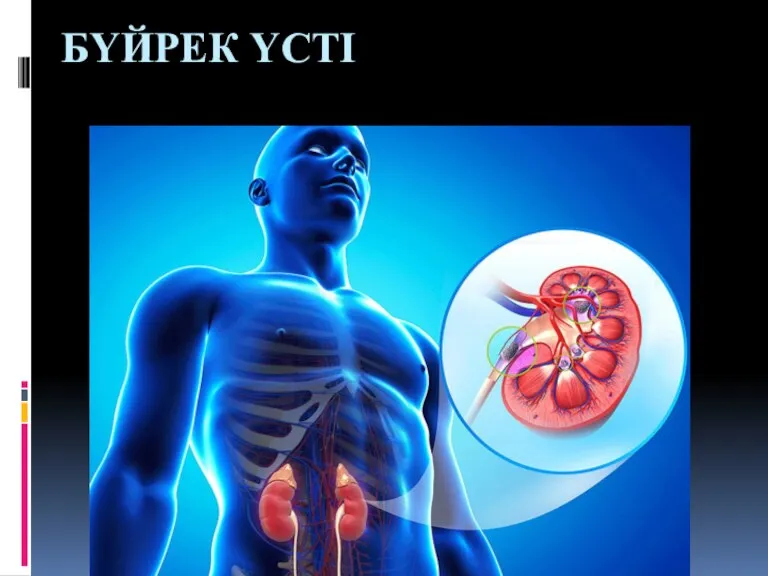 Бүйрек үсті
 Здоровое питание
 Терминальное состояние:стадии, клиника, диагностика, критерии оценки тяжести состояния больного
 Доброкачественные заболевания матки и придатков
 Острые отравления в педиатрической практике
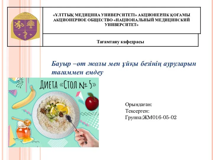 Бауыр –өт жолы мен ұйқы безінің. ауруларын тағаммен емдеу
 Первичный туберкулез у детей
 Диагностика и дифференциальная диагностика бруцеллеза и инфекционного эпидидимита баранов
 Жүрек аускультациясы. Жүрек тондары: қалыпты және патологиялық. Жүрек шулары: органикалық және функциональды шулар
 Ревматизм. Роль медсестры в лечении и профилактике
 Всемирная организация здравоохранения
 Инфузионная терапия и парентеральное питание у детей
 Медикаментозное лечение. Пути введения лекарственных средств. Способы применения лекарственных средств
 Наркоз. Стадии наркоза
 Сравнение функциональных возможностей статистических пакетов Мicrosoft office excel и Биостат
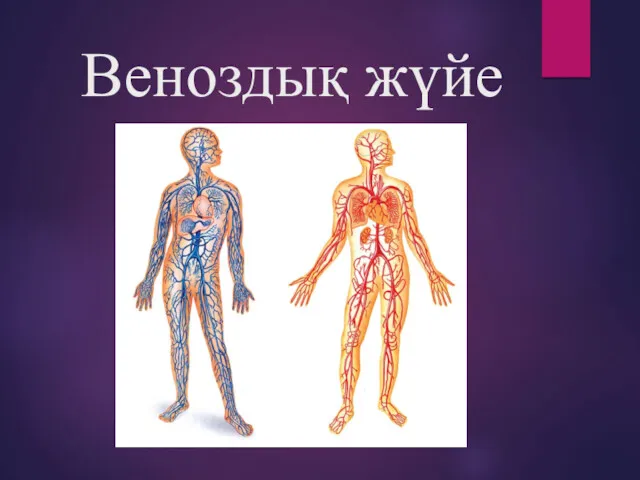 Веноздық жүйе
 Психофизиология. Главные задачи психофизиологии
 Искусственное кровообращение