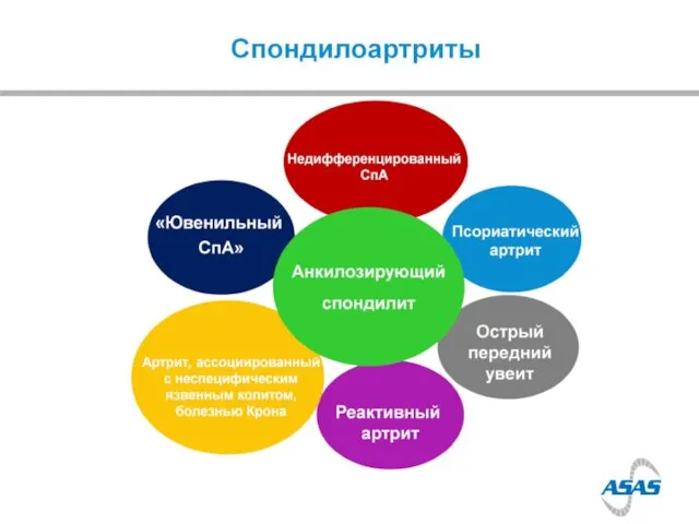
- Ankylosing Spondylitis

Содержание
- 3. Discovery 1890s Bechterew W. (1893). "Steifigkeit der Wirbelsaule und ihre Verkrummung als besondere Erkrankungsform". Neurol Centralbl
- 4. Description 20th century
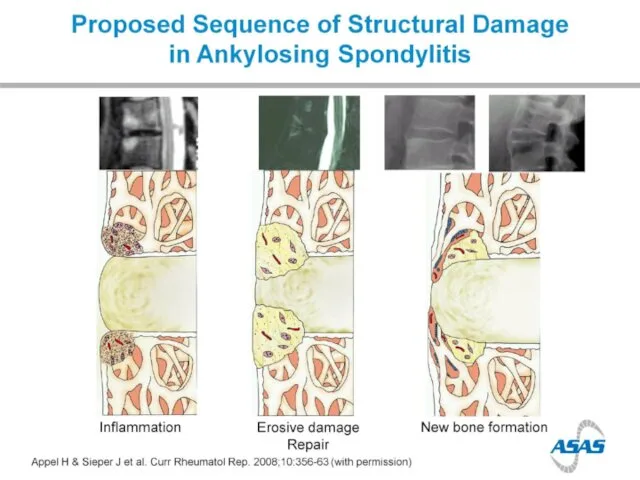
- 5. Bone formation in AS
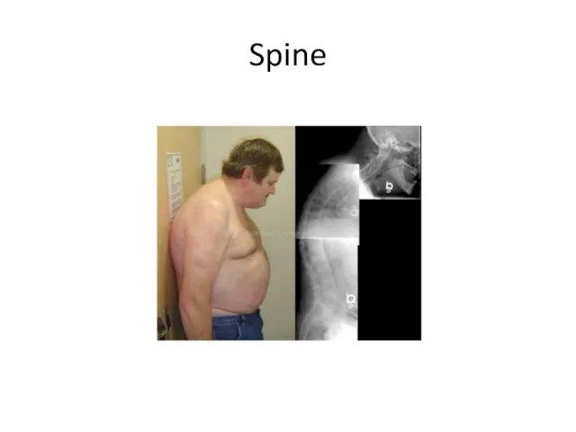
- 7. Spine
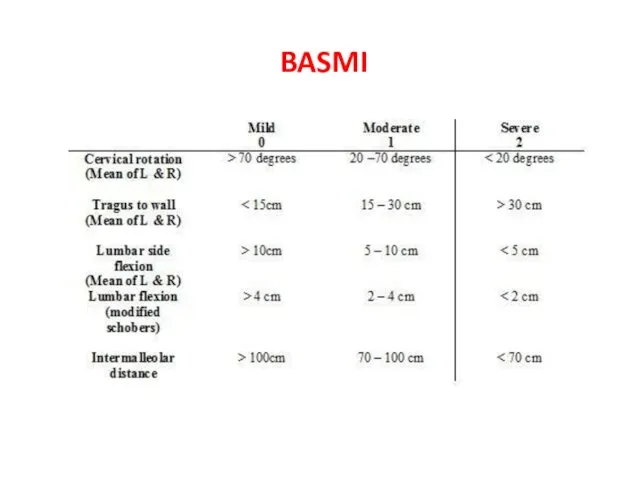
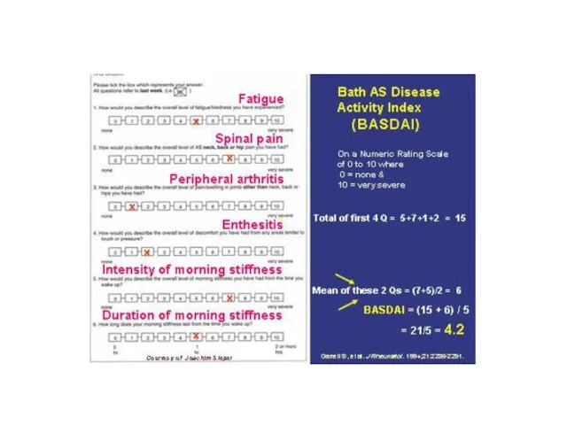
- 8. BASMI
- 10. Sacroiliac joints
- 11. Spine
- 16. Patient’s symptoms One half comes from inflammation, the second half comes from structural changes
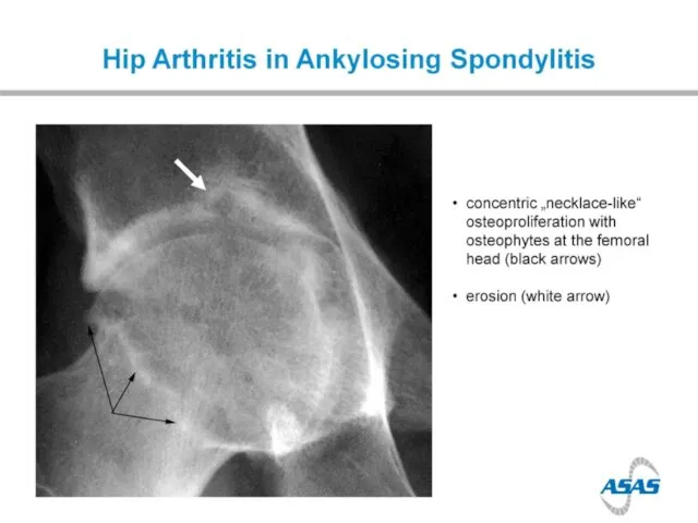
- 17. Arthritis
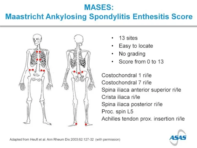
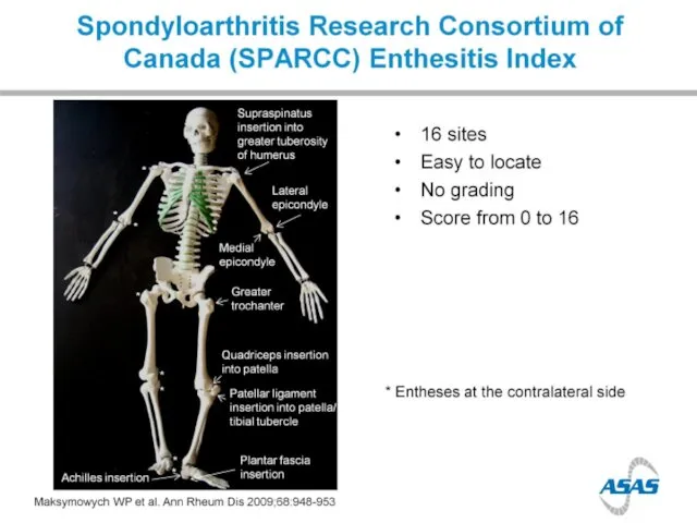
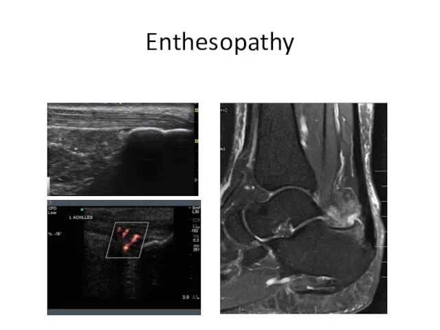
- 19. Enthesopathy
- 22. Enthesopathy
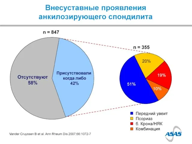
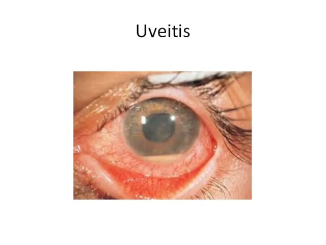
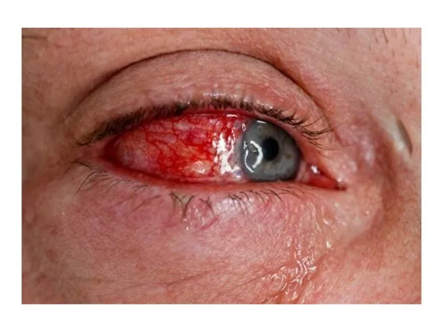
- 24. Uveitis
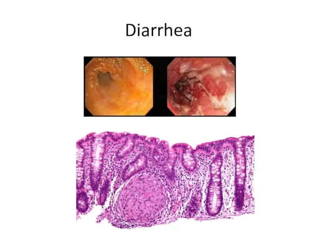
- 25. Diarrhea
- 27. Aortitis
- 29. HLA B27 1970s HLA B27 + in 90% of Caucasian AS pt Only 5% of HLA
- 35. Non X-ray imaging Computed tomography Better comparing to X-rays But not validated and radiation-related
- 36. Non X-ray imaging of SIJ Bone scan Sensitivity is about 50% Specificity is about 50%
- 37. Non X-ray imaging of SIJ MRI 22 yo F with right LBP
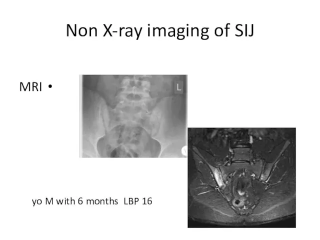
- 38. Non X-ray imaging of SIJ MRI 16 yo M with 6 months LBP
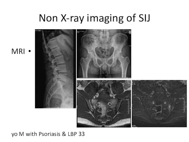
- 39. Non X-ray imaging of SIJ MRI 33 yo M with Psoriasis & LBP
- 40. Spine - MRI
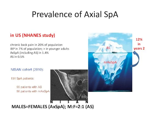
- 43. Prevalence of Axial SpA in US (NHANES study) chronic back pain in 20% of population IBP
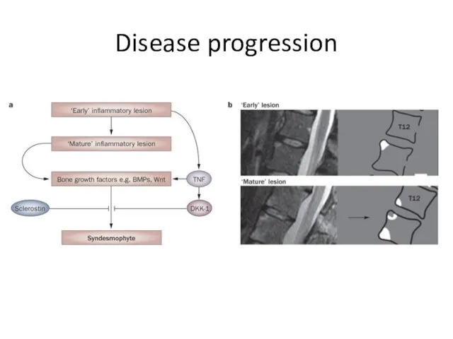
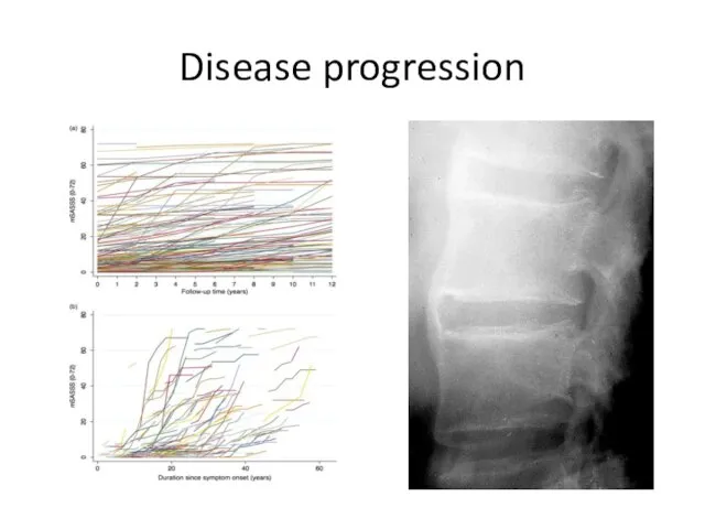
- 44. Disease progression
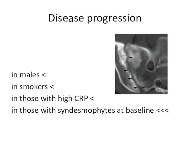
- 45. Disease progression > in males > in smokers > in those with high CRP >>> in
- 46. Disease progression
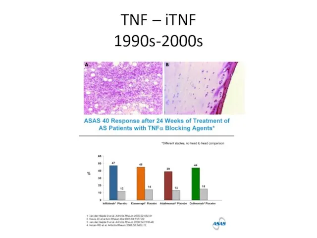
- 49. TNF – iTNF 1990s-2000s
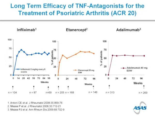
- 50. Efficacy of TNFi in AS
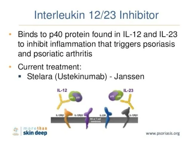
- 51. Other biologics: anti IL-17
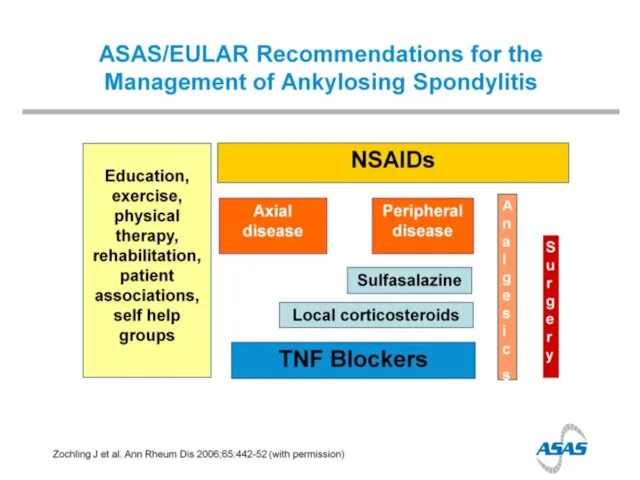
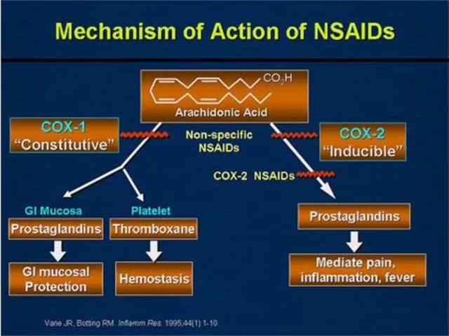
- 52. Anti-TNF & NSAIDS
- 53. A ROYAL FAMILY of rheumatic diseases
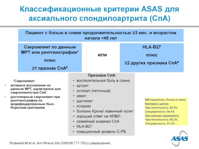
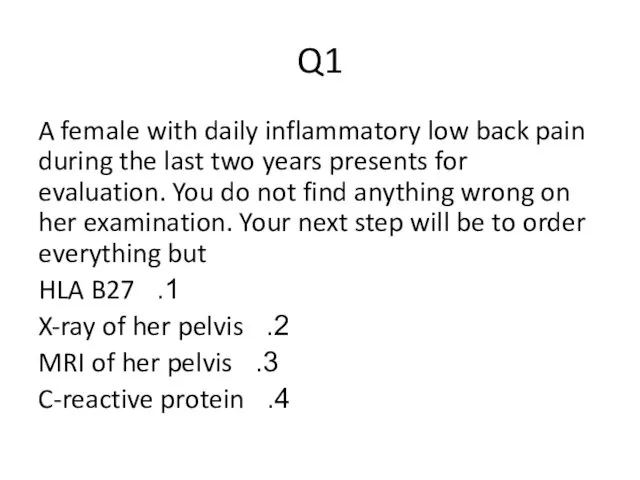
- 54. Q1 A female with daily inflammatory low back pain during the last two years presents for
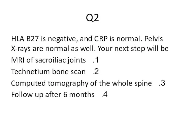
- 55. Q2 HLA B27 is negative, and CRP is normal. Pelvis X-rays are normal as well. Your
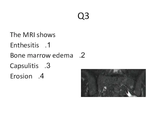
- 56. Q3 The MRI shows Enthesitis Bone marrow edema Capsulitis Erosion
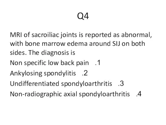
- 57. Q4 MRI of sacroiliac joints is reported as abnormal, with bone marrow edema around SIJ on
- 58. Q5 Non-radiographic axial spondyloarthritis is An early phase of ankylosing spondylitis A variant of osteitis condensanse
- 59. Q6 Recommended treatment will be NSAIDs Physical therapy TNF-alpha blockade Surgery
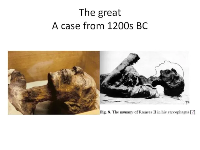
- 60. The great A case from 1200s BC
- 61. The great A case from 1200s BC
- 62. DISH vs AS
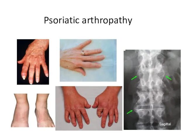
- 63. Psoriatic arthropathy
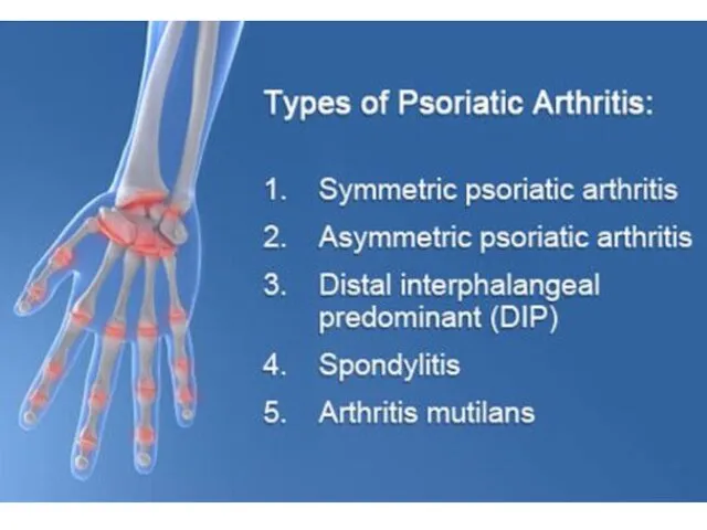
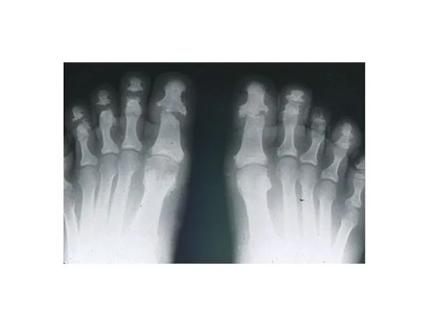
- 64. Psoriatic arthropathy
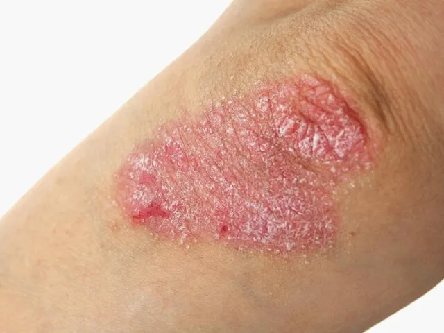
- 67. Psoriasis
- 68. Enthesopathy
- 69. Uveitis
- 71. Treatment
- 74. Reactive arthritis
- 78. Скачать презентацию
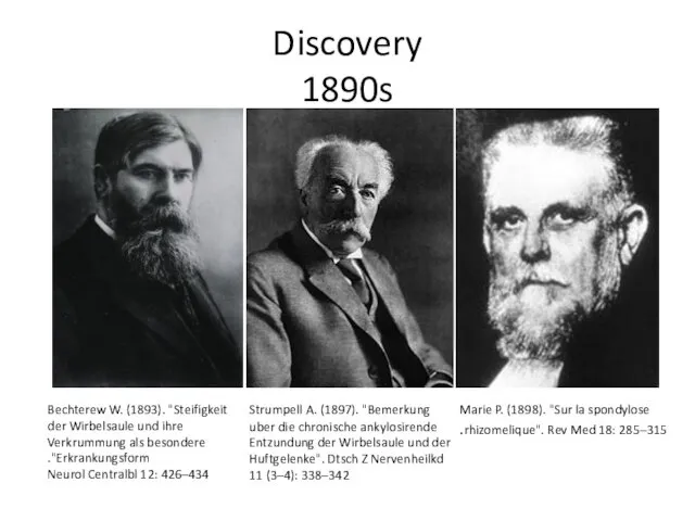
Discovery
1890s
Bechterew W. (1893). "Steifigkeit der Wirbelsaule und ihre Verkrummung als besondere
Discovery
1890s
Bechterew W. (1893). "Steifigkeit der Wirbelsaule und ihre Verkrummung als besondere
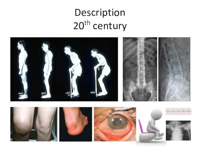
Description
20th century
Description
20th century
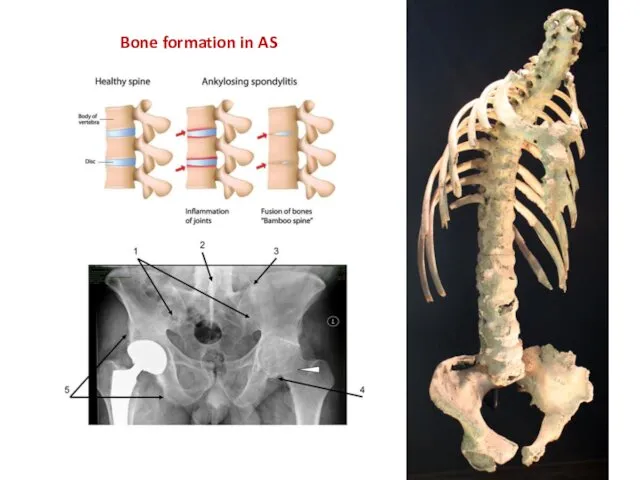
Bone formation in AS
Bone formation in AS
Spine
Spine
BASMI
BASMI
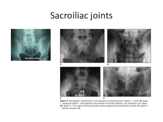
Sacroiliac joints
Sacroiliac joints
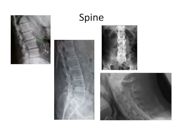
Spine
Spine
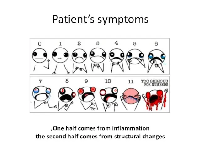
Patient’s symptoms
One half comes from inflammation,
the second half comes from
Patient’s symptoms
One half comes from inflammation,
the second half comes from
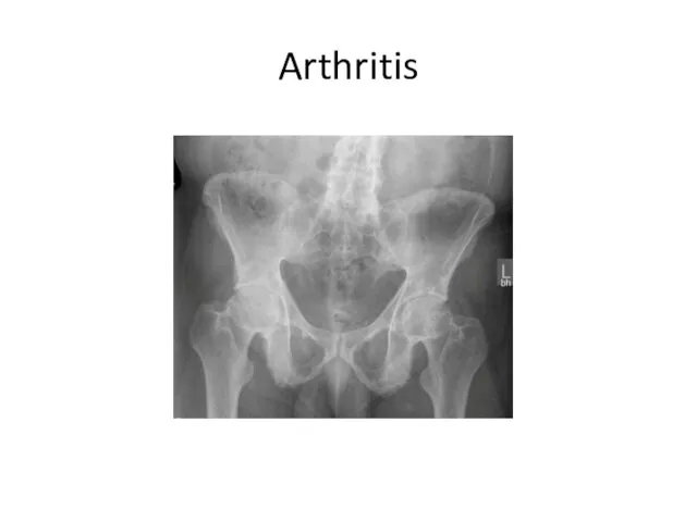
Arthritis
Arthritis
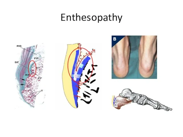
Enthesopathy
Enthesopathy
Enthesopathy
Enthesopathy
Uveitis
Uveitis
Diarrhea
Diarrhea
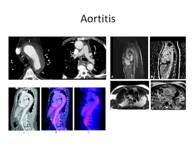
Aortitis
Aortitis
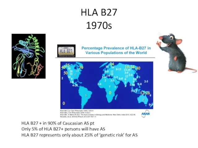
HLA B27
1970s
HLA B27 + in 90% of Caucasian AS pt
Only 5%
HLA B27
1970s
HLA B27 + in 90% of Caucasian AS pt
Only 5%
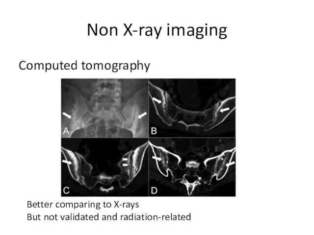
Non X-ray imaging
Computed tomography
Better comparing to X-rays
But not validated and
Non X-ray imaging
Computed tomography
Better comparing to X-rays
But not validated and
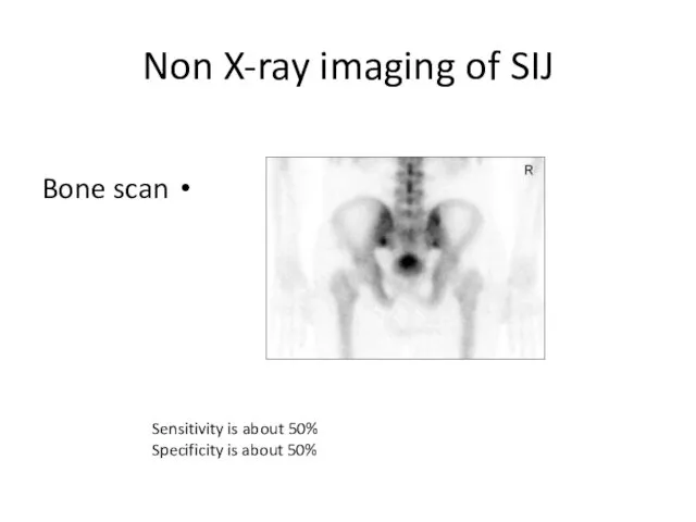
Non X-ray imaging of SIJ
Bone scan
Sensitivity is about 50%
Specificity is about
Non X-ray imaging of SIJ
Bone scan
Sensitivity is about 50%
Specificity is about
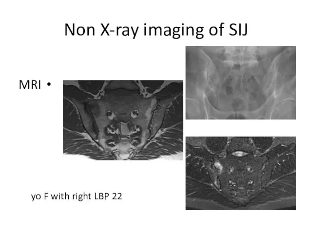
Non X-ray imaging of SIJ
MRI
22 yo F with right LBP
Non X-ray imaging of SIJ
MRI
22 yo F with right LBP
Non X-ray imaging of SIJ
MRI
16 yo M with 6 months LBP
Non X-ray imaging of SIJ
MRI
16 yo M with 6 months LBP
Non X-ray imaging of SIJ
MRI
33 yo M with Psoriasis & LBP
Non X-ray imaging of SIJ
MRI
33 yo M with Psoriasis & LBP
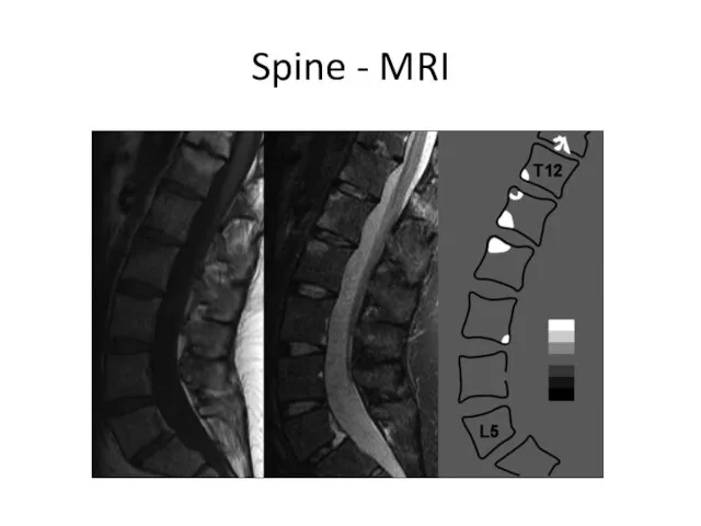
Spine - MRI
Spine - MRI
Prevalence of Axial SpA
in US (NHANES study)
chronic back pain in 20%
Prevalence of Axial SpA
in US (NHANES study)
chronic back pain in 20%
Disease progression
Disease progression
Disease progression
> in males
> in smokers
> in those with high CRP
>>>
Disease progression
> in males
> in smokers
> in those with high CRP
>>>
Disease progression
Disease progression
TNF – iTNF
1990s-2000s
TNF – iTNF
1990s-2000s
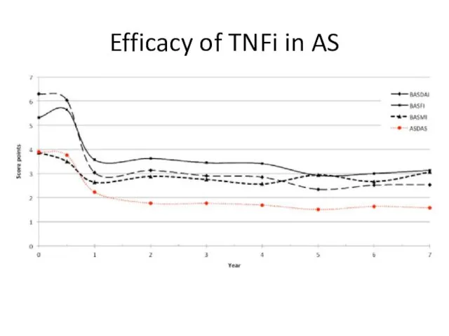
Efficacy of TNFi in AS
Efficacy of TNFi in AS
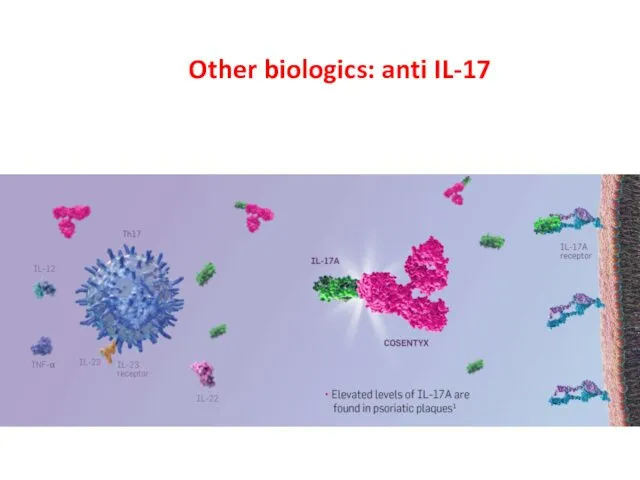
Other biologics: anti IL-17
Other biologics: anti IL-17
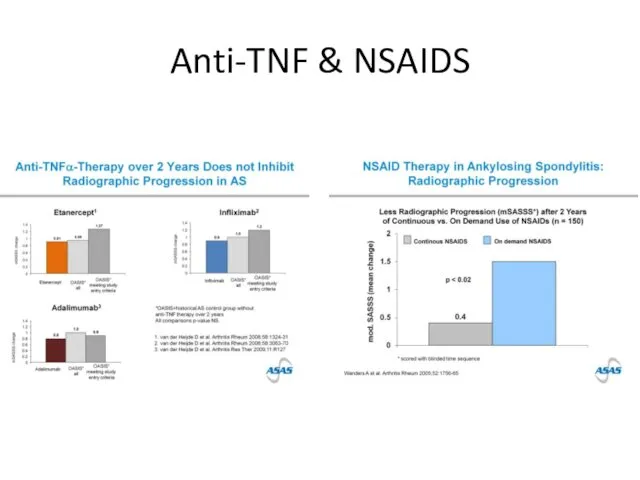
Anti-TNF & NSAIDS
Anti-TNF & NSAIDS
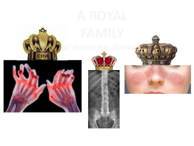
A ROYAL FAMILY
of rheumatic diseases
A ROYAL FAMILY
of rheumatic diseases
Q1
A female with daily inflammatory low back pain during the last
Q1
A female with daily inflammatory low back pain during the last
Q2
HLA B27 is negative, and CRP is normal. Pelvis X-rays are
Q2
HLA B27 is negative, and CRP is normal. Pelvis X-rays are
Q3
The MRI shows
Enthesitis
Bone marrow edema
Capsulitis
Erosion
Q3
The MRI shows
Enthesitis
Bone marrow edema
Capsulitis
Erosion
Q4
MRI of sacroiliac joints is reported as abnormal, with bone marrow
Q4
MRI of sacroiliac joints is reported as abnormal, with bone marrow
Q5
Non-radiographic axial spondyloarthritis is
An early phase of ankylosing spondylitis
A variant
Q5
Non-radiographic axial spondyloarthritis is
An early phase of ankylosing spondylitis
A variant

Q6
Recommended treatment will be
NSAIDs
Physical therapy
TNF-alpha blockade
Surgery
Q6
Recommended treatment will be
NSAIDs
Physical therapy
TNF-alpha blockade
Surgery
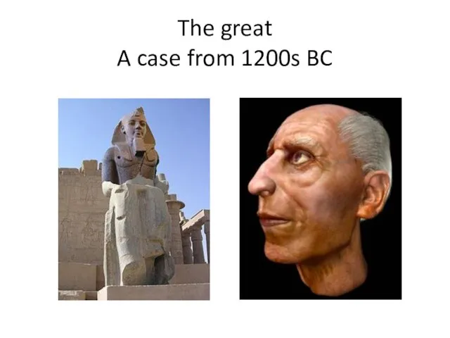
The great
A case from 1200s BC
The great
A case from 1200s BC
The great
A case from 1200s BC
The great
A case from 1200s BC
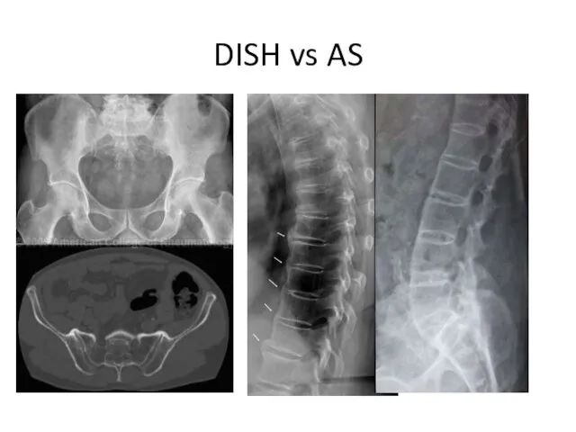
DISH vs AS
DISH vs AS
Psoriatic arthropathy
Psoriatic arthropathy
Psoriatic arthropathy
Psoriatic arthropathy
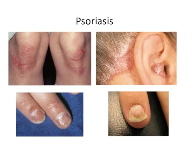
Psoriasis
Psoriasis
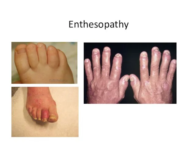
Enthesopathy
Enthesopathy
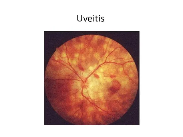
Uveitis
Uveitis
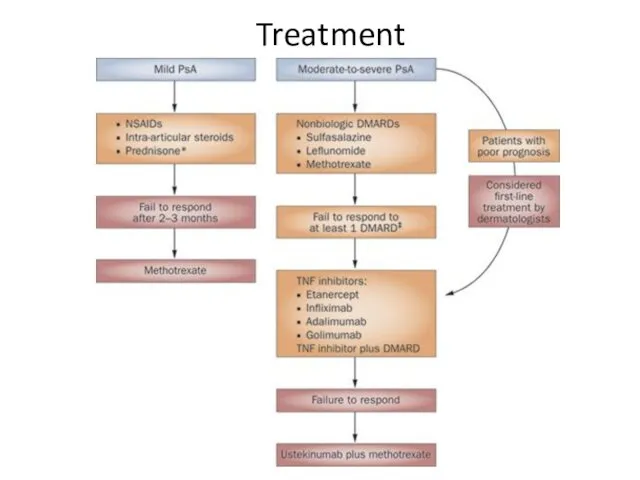
Treatment
Treatment
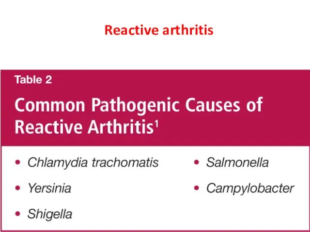
Reactive arthritis
Reactive arthritis

 Diabetes mellitus. (Subject 8)
 Омолаживающие операции на лице и шее
 Переход к системе непрерывного медицинского (фармацевтического) образования
 Контрацепция для подростков
 Правовая и юридическая база сестринского дела
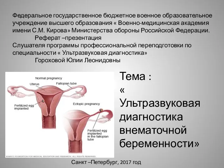 Ультразвуковая диагностика внематочной беременности
 Нәрестелер иммунитетіндегі иммуноглобулиндердің ролі
 Физиотерапия, кинезитерапия при травматических повреждениях
 Повреждение ключицы. Лечение
 Рентгенодиагностика при хроническом бруцеллезе
 Клубные наркотики
 Современное представление о нормальных родах
 Клинико-фармакологические подходы к выбору и применению антиаритмических лекарственных средств
 Бронхиальная астма
 Первая медицинская помощь при травмах
 Роль медицинской сестры при уходе за пациентами с сахарным диабетом II типа. Участие в лечебно-диагностических процессах
 Врачебная этика И. Канта
 Хроническое воспаление. Гранулематозное и специфическое воспаление
 Лечение ХОБЛ и бронхиальной астмы
 Чипсы: польза или вред
 Ошибки лабораторной диагностики. Нормативно-правовая база
 Клиника интеллектуальных нарушений при деменции
 Введение в эпидемиологию. Основы учения об эпидемическом процессе. Место эпидемиологии в структуре медицинских наук
 Профессиональное здоровье педагогов по результатам социологического опроса
 Turner syndrome
 Недоношенные новорожденные
 Лечение артериальной гипертензии в сочетании с сахарным диабетом
 Бүйрек-тас ауруы