- Syndrome of acute inflammation of mucous membranes of respiratory tracts. Tonsillitises

Содержание
- 2. SYNDROME OF ACUTE INFLAMMATION OF MUCOUS MEMBRANES of RESPIRATORY TRACTS Among diseases developing with inflammation of
- 3. Term «acute respiratory viral infections» (ARVI) is a group of viral diseases without concrete nosology. ARD
- 4. ARD is most widespread diseases. Annually over 25% of population is sick and morbidity rises considerably
- 5. To find out etiology. If etiology is not succeeded a diagnosis is formulated as «ARD undifferentiated»
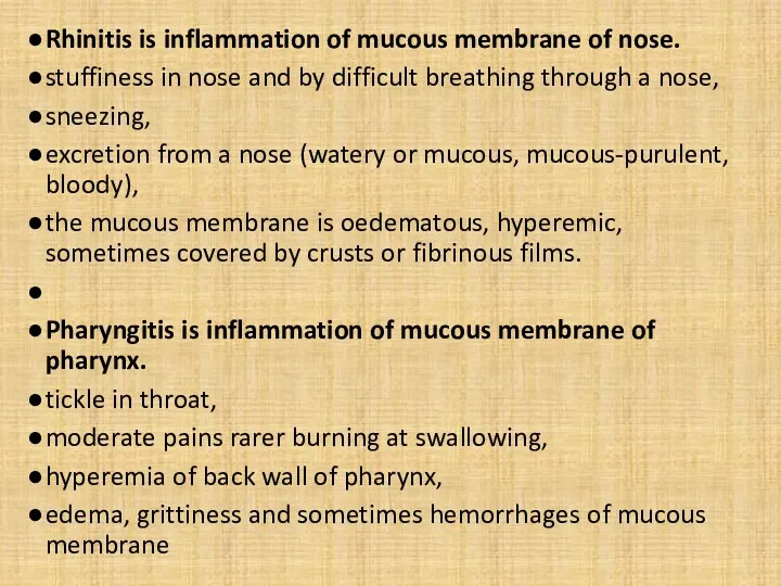
- 6. Rhinitis is inflammation of mucous membrane of nose. stuffiness in nose and by difficult breathing through
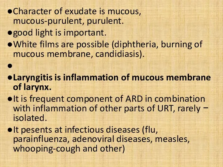
- 7. Character of exudate is mucous, mucous-purulent, purulent. good light is important. White films are possible (diphtheria,
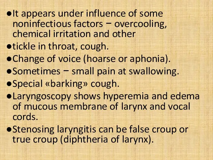
- 8. It appears under influence of some noninfectious factors − overcooling, chemical irritation and other tickle in
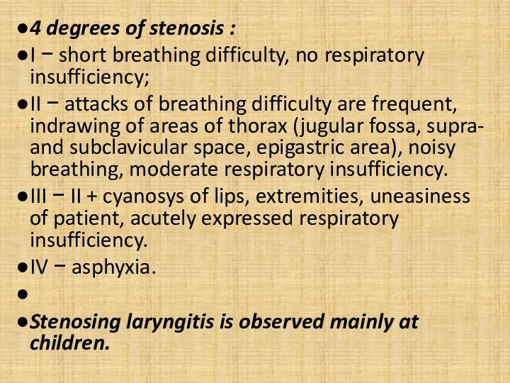
- 9. 4 degrees of stenosis : I − short breathing difficulty, no respiratory insufficiency; II − attacks
- 10. Chronic laryngitis at noninfectious pathology − hoarseness and rapid fatigueability of voice, tickle in throat, dryness.
- 11. As a component of ARVI if it combines with the damage of URT. For ARD an
- 12. A bronchiolitis is more severe form of acute bronchitis. Bronchioles are involved in a process. The
- 13. Next groups of diseases with inflammatory changes of upper respiratory tracts are possible: ARD; inflammation of
- 14. Acute respiratory diseases: adenoviral diseasees; bacterial nasopharyngitis (streptococcus, staphylococcus and other); herpetic respiratory diseasees; flu; coronaviral
- 15. respiratory syncytial infection (RS-virus disease); rhinovirus disease; enterovirus nasopharyngitis; mycoplasm inflammation of upper respiratory tracts; parainfluenza;
- 16. Infectious diseases developing with the signs of inflammation of respiratory tracts: herpangina; diphtheria; whooping-cough; measles; rubella;
- 17. Toxicochemical inflammatory changes of URT : develops without the expressed fever and signs of general intoxication;
- 18. Flu. A diagnosis is not difficult during epidemics. At the beginning of epidemic flu develops more
- 19. General intoxication is expressed (meningism, encephalopathy ). Hypotension. 85% cases last 3-5 days. The presence of
- 20. Parainfluenza Unlike a flu does not have such clear clinical presentation. There are not large epidemics.
- 21. The temperature of body, as a rule, is subfebrile. The symptoms of general intoxication are expressed
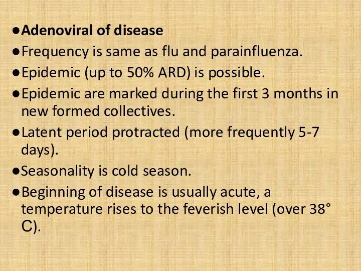
- 22. Adenoviral of disease Frequency is same as flu and parainfluenza. Epidemic (up to 50% ARD) is
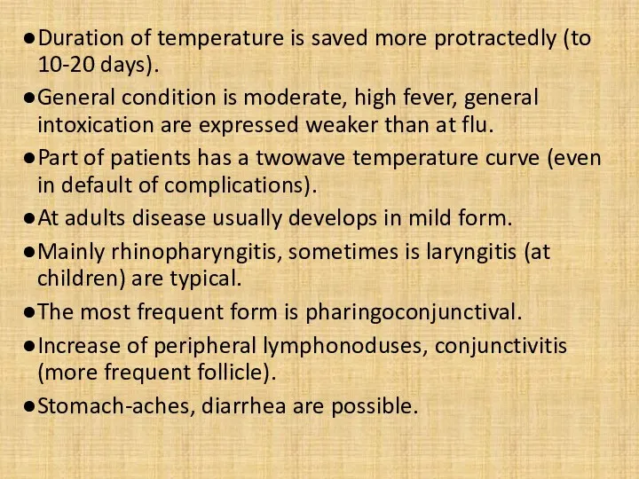
- 23. Duration of temperature is saved more protractedly (to 10-20 days). General condition is moderate, high fever,
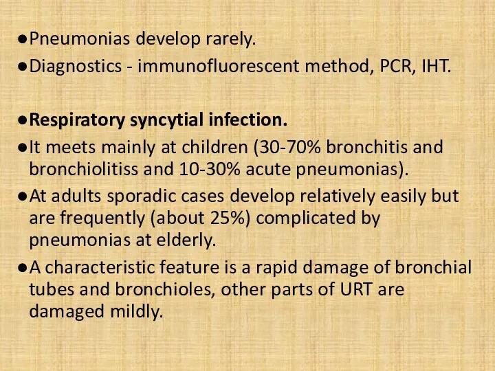
- 24. Pneumonias develop rarely. Diagnostics - immunofluorescent method, PCR, IHT. Respiratory syncytial infection. It meets mainly at
- 25. Development is more protracted than at other ARD. Diagnostics - immunofluorescent method, PCR, IHT. Rhinovirus disease.
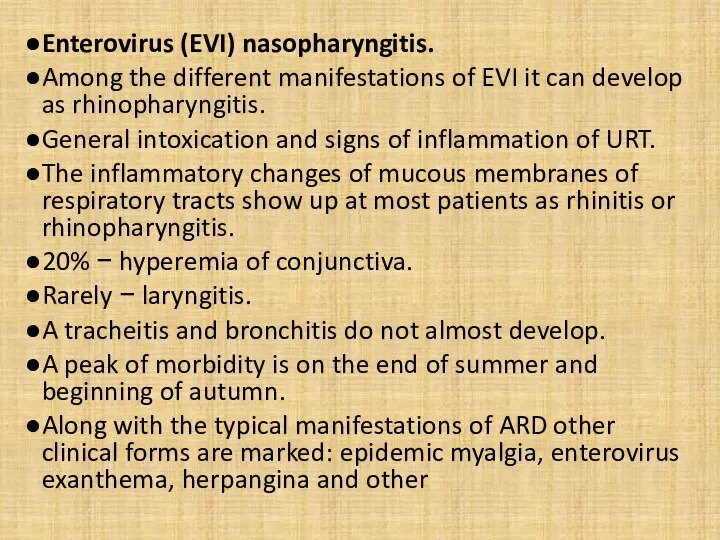
- 26. Enterovirus (EVI) nasopharyngitis. Among the different manifestations of EVI it can develop as rhinopharyngitis. General intoxication
- 27. These manifestations are original indicators of EVI. Sporadic cases is difficult to recognize clinically. Diagnostics -
- 28. Mycoplasm inflammation of upper respiratory tracts. М. рпеитоniе, experiments on volunteers show possibility of exsudative pharyngitis
- 29. Bacterial ARD. Streptococci, staphylococci and other cause isolated damages of some part of URT. Antibiotic therapy
- 30. Streptococcal pharyngitis. Вeta-haemolytic streptococcustococcus group А is characteristic for pharyngitis and other streptococcus diseases (scarlatina, quinsies,
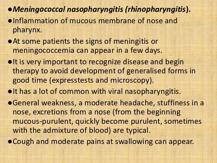
- 31. Meningococcal nasopharyngitis (rhinopharyngitis). Inflammation of mucous membrane of nose and pharynx. At some patients the signs
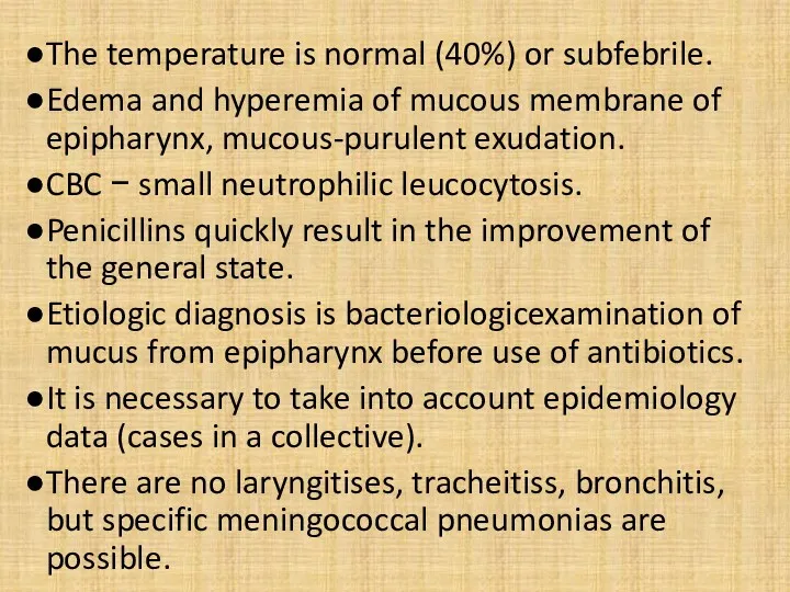
- 32. The temperature is normal (40%) or subfebrile. Edema and hyperemia of mucous membrane of epipharynx, mucous-purulent
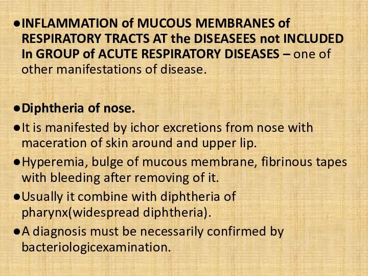
- 33. INFLAMMATION of MUCOUS MEMBRANES of RESPIRATORY TRACTS AT the DISEASEES not INCLUDED In GROUP of ACUTE
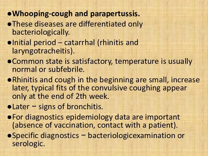
- 34. Whooping-cough and parapertussis. These diseases are differentiated only bacteriologically. Initial period – catarrhal (rhinitis and laryngotracheitis).
- 35. Measles. Before appearance of characteristic exanthema it is frequently interpreted as ARD. Fever, pharyngitis. Expressed conjunctivitis
- 36. Rubella. Signs of acute rhinopharyngitis, small hyperemia of conjunctiva, subfebrile temperature of body are possible at
- 37. Herpangina. Pharyngitis, intoxication. Enanthema: hyperemia − small red spots (2-3 mm) - vesiculas (4-5 mm) -
- 38. Q-Fever. Bronchial tubes are damaged mainly, but moderate inflammation of upper respiratory tracts as pharyngitis and
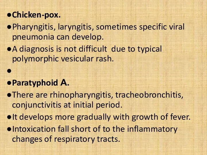
- 39. Chicken-pox. Pharyngitis, laryngitis, sometimes specific viral pneumonia can develop. A diagnosis is not difficult due to
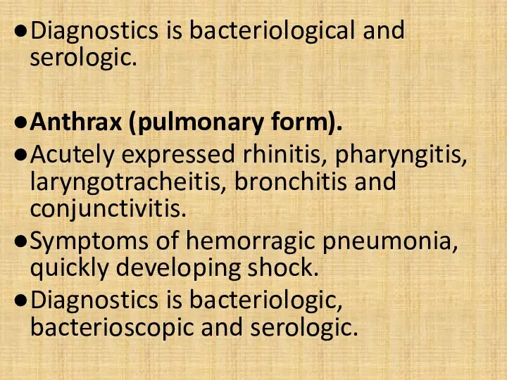
- 40. Diagnostics is bacteriological and serologic. Anthrax (pulmonary form). Acutely expressed rhinitis, pharyngitis, laryngotracheitis, bronchitis and conjunctivitis.
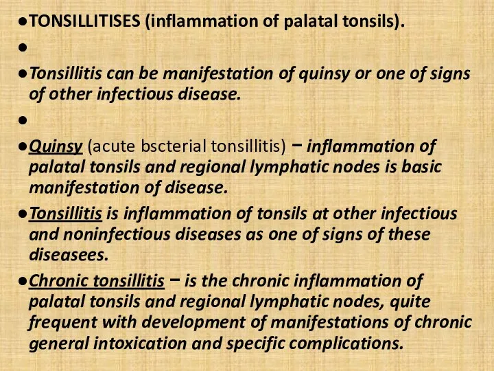
- 41. TONSILLITISES (inflammation of palatal tonsils). Tonsillitis can be manifestation of quinsy or one of signs of
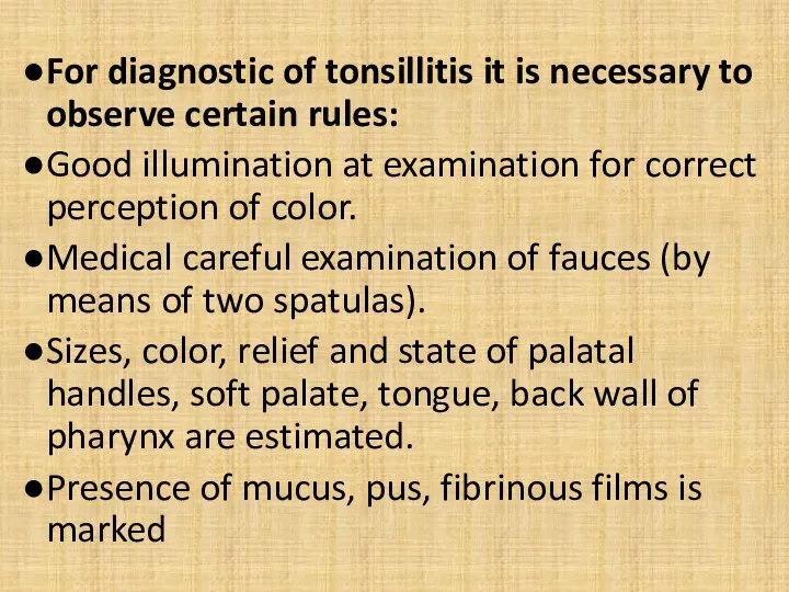
- 42. For diagnostic of tonsillitis it is necessary to observe certain rules: Good illumination at examination for
- 43. Quinsy. It can be caused by haemolytic streptococcus (85%), staphylococcus aureus (10%), fusobacteria (necrotic Vincent's quinsy).
- 44. Features of necrotic Vincent's quinsy: absence of the expressed symptoms of general intoxication, pharyngalgia at swallowing
- 45. Infectious: adenoviral diseases; anginal-bubonic form of rabbit-fever; anginal-septic form of listeriosis; diphtheria of pharynx; infectious mononucleosis;
- 46. Noninfectious: agranulocytosis radiation disease; acute leucosises; cytostatic disease; chronic tonsillitis.
- 47. Adenoviral of disease. Adenoviruss can save long time in tissue of tonsils and adenoids without inflammatory
- 48. Film on the tonsil has greyish color and deep ulcer appears after sloughing. After repairing of
- 49. Anginal-septic form of listeriosis. Tonsillitis develops on a background a severe general (septic) disease with the
- 50. Diagnostics − bacteriologic (from blood, neurolymph, pharynx), serologic (CFR and IHR). A listerious allergen is produced
- 51. Catarrhal form of diphtheria of pharynx. Atypical form, without fibrinous films on tonsils. The temperature of
- 52. Island-like form of diphtheria of pharynx. The small areas of fibrinousого film (small «islands») on tonsils
- 53. Membranous diphtheria of pharynx. Expressed continuous fibrinous films keeping indoors outside tonsils. A film is taken
- 54. Toxic diphtheria is characterized by appearance of edema of neck cellular tissue. At toxic diphtheria false
- 55. Appearance of paratonsillar abscess is characterized by a rapid fervescence, chill expressed by a toxicosis. Trismus
- 56. Appearance of tonsillitis is possible after ferver, general intoxication and other signs of mononucleosis. It can
- 57. Acute hepatitis can develop. CBC − leucocytosis, neutropenia, lymphomonocytosis, atypical mononuclears. The changes of blood appear
- 58. Candidiasis of fauces. Arises up as a result of dysbacteriosis and immunodeficit. The temperature of body
- 59. Inflammation of tonsils is not actual it is not tonsillitis but widespread candida damage of mucous
- 60. A regional lymphatic node is mildly increased, dense consistency, painless. Pains at swallowing is not marked.
- 61. Scarlatina Like quinsy it is caused by haemolytic streptococcustococcus group A. Along with a clinic signs
- 62. Typhoid fever. Palatal tonsils and archs are edematous, small ulcers covered by a greyish films are
- 63. Chronic tonsillitis. Character of development is decompensated and subcompensated. Periodic exacerbation and remissions are typical. Exacerbation
- 65. Скачать презентацию
SYNDROME OF ACUTE INFLAMMATION OF MUCOUS MEMBRANES of RESPIRATORY TRACTS
Among diseases
SYNDROME OF ACUTE INFLAMMATION OF MUCOUS MEMBRANES of RESPIRATORY TRACTS
Among diseases
Term «acute respiratory viral infections» (ARVI) is a group of viral
Term «acute respiratory viral infections» (ARVI) is a group of viral
ARD is most widespread diseases.
Annually over 25% of population is
ARD is most widespread diseases.
Annually over 25% of population is
To find out etiology.
If etiology is not succeeded a diagnosis
To find out etiology.
If etiology is not succeeded a diagnosis
Rhinitis is inflammation of mucous membrane of nose.
stuffiness in nose and
Rhinitis is inflammation of mucous membrane of nose.
stuffiness in nose and
Character of exudate is mucous, mucous-purulent, purulent.
good light is important.
White
Character of exudate is mucous, mucous-purulent, purulent.
good light is important.
White
It appears under influence of some noninfectious factors − overcooling, chemical
It appears under influence of some noninfectious factors − overcooling, chemical
4 degrees of stenosis :
I − short breathing difficulty, no
4 degrees of stenosis :
I − short breathing difficulty, no
Chronic laryngitis at noninfectious pathology − hoarseness and rapid fatigueability of
Chronic laryngitis at noninfectious pathology − hoarseness and rapid fatigueability of
As a component of ARVI if it combines with the damage
As a component of ARVI if it combines with the damage
A bronchiolitis is more severe form of acute bronchitis.
Bronchioles are involved
A bronchiolitis is more severe form of acute bronchitis.
Bronchioles are involved
Next groups of diseases with inflammatory changes of upper respiratory tracts
Next groups of diseases with inflammatory changes of upper respiratory tracts
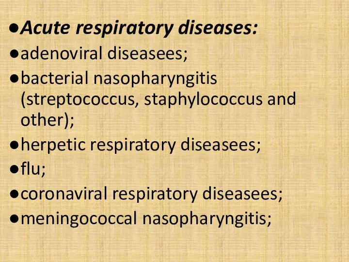
Acute respiratory diseases:
adenoviral diseasees;
bacterial nasopharyngitis (streptococcus, staphylococcus and other);
herpetic respiratory diseasees;
flu;
coronaviral
Acute respiratory diseases:
adenoviral diseasees;
bacterial nasopharyngitis (streptococcus, staphylococcus and other);
herpetic respiratory diseasees;
flu;
coronaviral
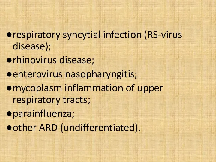
respiratory syncytial infection (RS-virus disease);
rhinovirus disease;
enterovirus nasopharyngitis;
mycoplasm inflammation of upper respiratory
respiratory syncytial infection (RS-virus disease);
rhinovirus disease;
enterovirus nasopharyngitis;
mycoplasm inflammation of upper respiratory
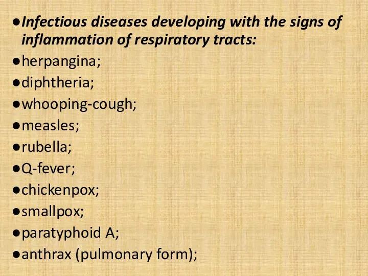
Infectious diseases developing with the signs of inflammation of respiratory tracts:
herpangina;
diphtheria;
whooping-cough;
measles;
rubella;
Q-fever;
chickenpox;
smallpox;
paratyphoid
Infectious diseases developing with the signs of inflammation of respiratory tracts:
herpangina;
diphtheria;
whooping-cough;
measles;
rubella;
Q-fever;
chickenpox;
smallpox;
paratyphoid
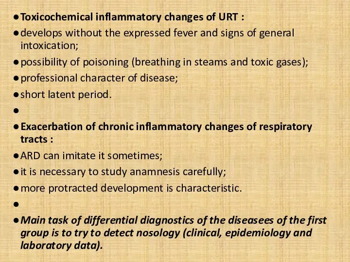
Toxicochemical inflammatory changes of URT :
develops without the expressed fever and
Toxicochemical inflammatory changes of URT :
develops without the expressed fever and
Flu. A diagnosis is not difficult during epidemics.
At the beginning of
Flu. A diagnosis is not difficult during epidemics.
At the beginning of
General intoxication is expressed (meningism, encephalopathy ).
Hypotension.
85% cases last 3-5
General intoxication is expressed (meningism, encephalopathy ).
Hypotension.
85% cases last 3-5
Parainfluenza
Unlike a flu does not have such clear clinical presentation.
Parainfluenza
Unlike a flu does not have such clear clinical presentation.
The temperature of body, as a rule, is subfebrile.
The symptoms of
The temperature of body, as a rule, is subfebrile.
The symptoms of
Adenoviral of disease
Frequency is same as flu and parainfluenza.
Epidemic (up to
Adenoviral of disease
Frequency is same as flu and parainfluenza.
Epidemic (up to
Duration of temperature is saved more protractedly (to 10-20 days).
General
Duration of temperature is saved more protractedly (to 10-20 days).
General
Pneumonias develop rarely.
Diagnostics - immunofluorescent method, PCR, IHT.
Respiratory syncytial infection.
It meets
Pneumonias develop rarely.
Diagnostics - immunofluorescent method, PCR, IHT.
Respiratory syncytial infection.
It meets

Development is more protracted than at other ARD.
Diagnostics - immunofluorescent method,
Development is more protracted than at other ARD.
Diagnostics - immunofluorescent method,
Enterovirus (EVI) nasopharyngitis.
Among the different manifestations of EVI it can
Enterovirus (EVI) nasopharyngitis.
Among the different manifestations of EVI it can
These manifestations are original indicators of EVI. Sporadic cases is difficult
These manifestations are original indicators of EVI. Sporadic cases is difficult
Mycoplasm inflammation of upper respiratory tracts.
М. рпеитоniе, experiments on volunteers
Mycoplasm inflammation of upper respiratory tracts.
М. рпеитоniе, experiments on volunteers
Bacterial ARD.
Streptococci, staphylococci and other cause isolated damages of some
Bacterial ARD.
Streptococci, staphylococci and other cause isolated damages of some
Streptococcal pharyngitis.
Вeta-haemolytic streptococcustococcus group А is characteristic for pharyngitis and
Streptococcal pharyngitis.
Вeta-haemolytic streptococcustococcus group А is characteristic for pharyngitis and
Meningococcal nasopharyngitis (rhinopharyngitis).
Inflammation of mucous membrane of nose and pharynx.
At
Meningococcal nasopharyngitis (rhinopharyngitis).
Inflammation of mucous membrane of nose and pharynx.
At
The temperature is normal (40%) or subfebrile.
Edema and hyperemia of
The temperature is normal (40%) or subfebrile.
Edema and hyperemia of
INFLAMMATION of MUCOUS MEMBRANES of RESPIRATORY TRACTS AT the DISEASEES not
INFLAMMATION of MUCOUS MEMBRANES of RESPIRATORY TRACTS AT the DISEASEES not
Whooping-cough and parapertussis.
These diseases are differentiated only bacteriologically.
Initial period
Whooping-cough and parapertussis.
These diseases are differentiated only bacteriologically.
Initial period
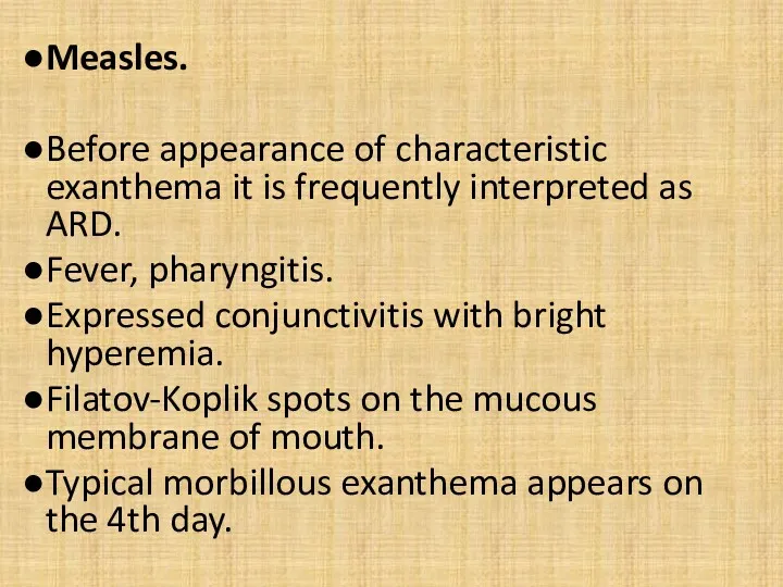
Measles.
Before appearance of characteristic exanthema it is frequently interpreted as
Measles.
Before appearance of characteristic exanthema it is frequently interpreted as
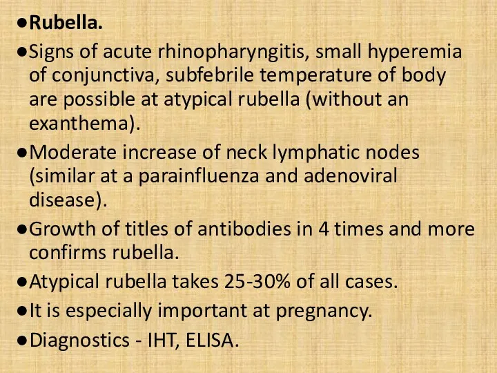
Rubella.
Signs of acute rhinopharyngitis, small hyperemia of conjunctiva, subfebrile temperature
Rubella.
Signs of acute rhinopharyngitis, small hyperemia of conjunctiva, subfebrile temperature
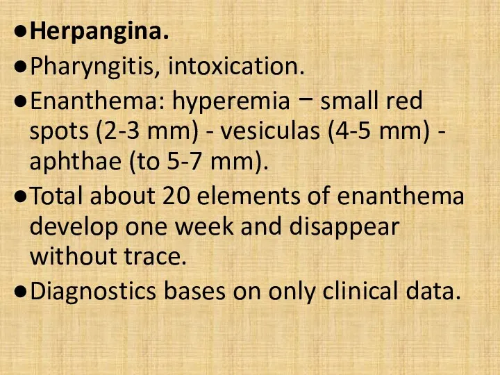
Herpangina.
Pharyngitis, intoxication.
Enanthema: hyperemia − small red spots (2-3 mm) -
Herpangina.
Pharyngitis, intoxication.
Enanthema: hyperemia − small red spots (2-3 mm) -
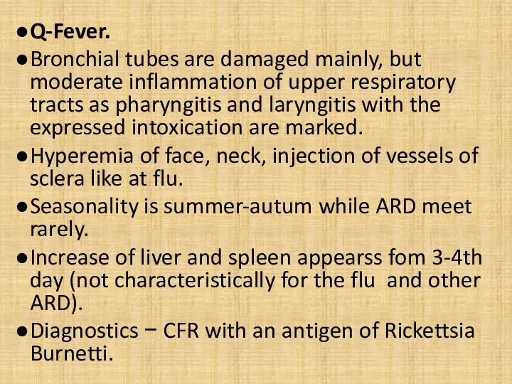
Q-Fever.
Bronchial tubes are damaged mainly, but moderate inflammation of upper
Q-Fever.
Bronchial tubes are damaged mainly, but moderate inflammation of upper
Chicken-pox.
Pharyngitis, laryngitis, sometimes specific viral pneumonia can develop.
A diagnosis
Chicken-pox.
Pharyngitis, laryngitis, sometimes specific viral pneumonia can develop.
A diagnosis
Diagnostics is bacteriological and serologic.
Anthrax (pulmonary form).
Acutely expressed rhinitis, pharyngitis,
Diagnostics is bacteriological and serologic.
Anthrax (pulmonary form).
Acutely expressed rhinitis, pharyngitis,
TONSILLITISES (inflammation of palatal tonsils).
Tonsillitis can be manifestation of quinsy or
TONSILLITISES (inflammation of palatal tonsils).
Tonsillitis can be manifestation of quinsy or
For diagnostic of tonsillitis it is necessary to observe certain rules:
Good
For diagnostic of tonsillitis it is necessary to observe certain rules:
Good
Quinsy.
It can be caused by haemolytic streptococcus (85%), staphylococcus aureus (10%),
Quinsy.
It can be caused by haemolytic streptococcus (85%), staphylococcus aureus (10%),
Features of necrotic Vincent's quinsy:
absence of the expressed symptoms of
Features of necrotic Vincent's quinsy:
absence of the expressed symptoms of
Infectious:
adenoviral diseases;
anginal-bubonic form of rabbit-fever;
anginal-septic form of listeriosis;
diphtheria of pharynx;
infectious mononucleosis;
candidiasis
Infectious:
adenoviral diseases;
anginal-bubonic form of rabbit-fever;
anginal-septic form of listeriosis;
diphtheria of pharynx;
infectious mononucleosis;
candidiasis
Noninfectious:
agranulocytosis
radiation disease;
acute leucosises;
cytostatic disease;
chronic tonsillitis.
Noninfectious:
agranulocytosis
radiation disease;
acute leucosises;
cytostatic disease;
chronic tonsillitis.
Adenoviral of disease.
Adenoviruss can save long time in tissue of
Adenoviral of disease.
Adenoviruss can save long time in tissue of
Film on the tonsil has greyish color and deep ulcer appears
Film on the tonsil has greyish color and deep ulcer appears
Anginal-septic form of listeriosis.
Tonsillitis develops on a background a severe general
Anginal-septic form of listeriosis.
Tonsillitis develops on a background a severe general
Diagnostics − bacteriologic (from blood, neurolymph, pharynx), serologic (CFR and IHR).
Diagnostics − bacteriologic (from blood, neurolymph, pharynx), serologic (CFR and IHR).
Catarrhal form of diphtheria of pharynx.
Atypical form, without fibrinous films
Catarrhal form of diphtheria of pharynx.
Atypical form, without fibrinous films
Island-like form of diphtheria of pharynx.
The small areas of fibrinousого film
Island-like form of diphtheria of pharynx.
The small areas of fibrinousого film
Membranous diphtheria of pharynx.
Expressed continuous fibrinous films keeping indoors outside tonsils.
A
Membranous diphtheria of pharynx.
Expressed continuous fibrinous films keeping indoors outside tonsils.
A
Toxic diphtheria is characterized by appearance of edema of neck cellular
Toxic diphtheria is characterized by appearance of edema of neck cellular
Appearance of paratonsillar abscess is characterized by a rapid fervescence, chill
Appearance of paratonsillar abscess is characterized by a rapid fervescence, chill
Appearance of tonsillitis is possible after ferver, general intoxication and
Appearance of tonsillitis is possible after ferver, general intoxication and
Acute hepatitis can develop.
CBC − leucocytosis, neutropenia, lymphomonocytosis, atypical mononuclears.
The
Acute hepatitis can develop.
CBC − leucocytosis, neutropenia, lymphomonocytosis, atypical mononuclears.
The
Candidiasis of fauces.
Arises up as a result of dysbacteriosis and
Candidiasis of fauces.
Arises up as a result of dysbacteriosis and
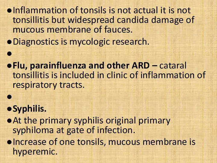
Inflammation of tonsils is not actual it is not tonsillitis but
Inflammation of tonsils is not actual it is not tonsillitis but
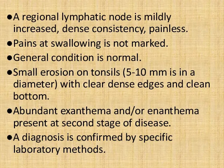
A regional lymphatic node is mildly increased, dense consistency, painless.
Pains
A regional lymphatic node is mildly increased, dense consistency, painless.
Pains
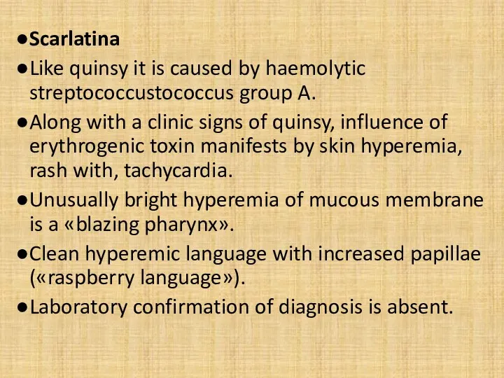
Scarlatina
Like quinsy it is caused by haemolytic streptococcustococcus group A.
Along
Scarlatina
Like quinsy it is caused by haemolytic streptococcustococcus group A.
Along
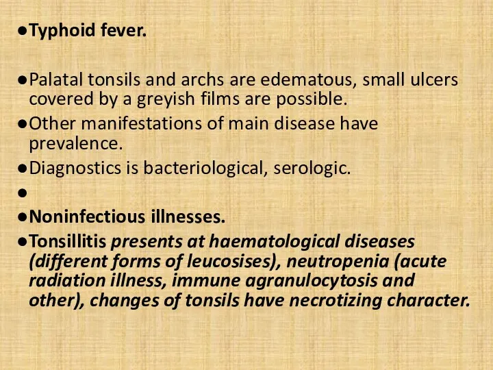
Typhoid fever.
Palatal tonsils and archs are edematous, small ulcers covered
Typhoid fever.
Palatal tonsils and archs are edematous, small ulcers covered
Chronic tonsillitis.
Character of development is decompensated and subcompensated.
Periodic exacerbation and
Chronic tonsillitis.
Character of development is decompensated and subcompensated.
Periodic exacerbation and
 Болезни желудка
 Детский массаж до года. (Занятие 2)
 Этиология и патогенез кариеса зубов. Современные представления
 Асқорыту мүшелеріне ауыз қуысы , өңеш, асқазан және ішек жатады. Асқорытуда ұйқы безі және бауыр қатысады
 Вирус иммунодефицита человека (ВИЧ)
 Заикание. Типы заикания. Диагностика. Лечение
 Абразивный преканцерозный хейлит Манганотти
 Проектная деятельность как средство формирования метапредметных умений на уроках математики
 Сәйкестендіру тесті
 Ротавирусы. Ротавирусный гастроэнтерит
 Дифференцировка Т-лимфоцитов
 Хирургиядағы ақпаратты–компьютерлік технологиялар, телемедицинасы
 Оценить эффективность комбинированной терапии (бетаблокатор+ингибитор апф) у пациентов в возрасте 18-55 лет с І и ІІ степенью АГ
 Твердые и мягкие лекарственные формы
 Жанұя денсаулығы. Бала денсаулығы
 Первая медицинская помощь при ДТП
 Здоровьесберегающие технологии профессора В.Ф. Базарного
 Варикозды кеңею. Тромбофлебит. Тромбофлебиттен кейінгі синдром
 Сахарный диабет
 Диссеминированный туберкулез легких
 Дәрігер-медбике-науқастың арасындағы қарым-қатынас
 Вирус иммунодефицита человека (ВИЧ). Вирус гриппа
 Жасөспірімдер мен балалар организіміне ішімдіктің әсерінің зияндылығын анализдеу
 Основы кардиологии мелких домашних животных для врачей общей практики
 Медикаментозное лечение. Пути введения лекарственных средств. Способы применения лекарственных средств
 Ішек биоценозы
 Холецистэктомия. Операция по удалению желчного пузыря
 Клиническая симптоматология ревматоидного артрита, остеоартроза