- The cardiac cycle and ECG

Содержание
- 2. The cardiac cycle and ECG
- 3. Learning objectives 11.1.3.4 use an electrocardiogram to describe the cardiac cycle 11.1.3.5 explain oxygen dissociation curves
- 4. Success creteria 1.Investigate the electrical process of in the heart . 2.Describe the structure of the
- 5. Terminology Atrium, ventricle, contract, systole, blood pressure, valves, aorta, pulmonary vein and arteria, relax, low and
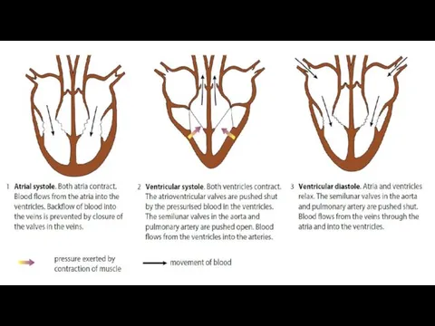
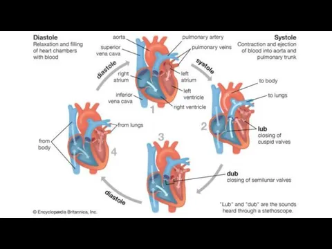
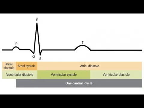
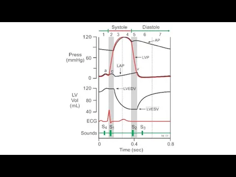
- 7. Cardiac cycle Your heart beats around 70 times a minute. The cardiac cycle is the sequence
- 8. Atrial systole The heart is filled with blood and the muscle in the atrial walls contracts.
- 9. Ventricular systole About 0.1 seconds after the atria contract, the ventricles contract. This is called ventricular
- 10. Ventricular diastole Ventricular systole lasts for about 0.3 seconds. The muscle then relaxes, and the stage
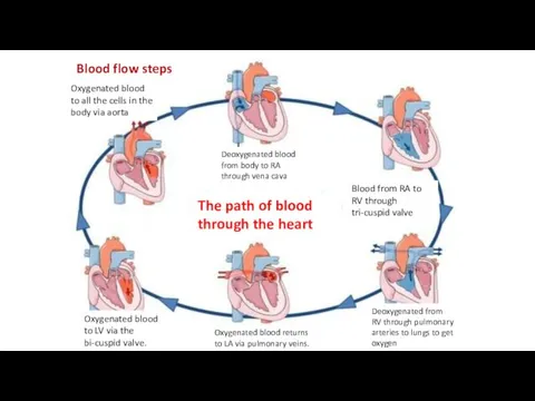
- 13. Deoxygenated blood from body to RA through vena cava Blood from RA to RV through tri-cuspid
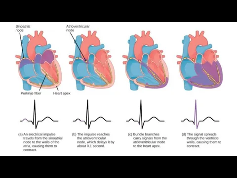
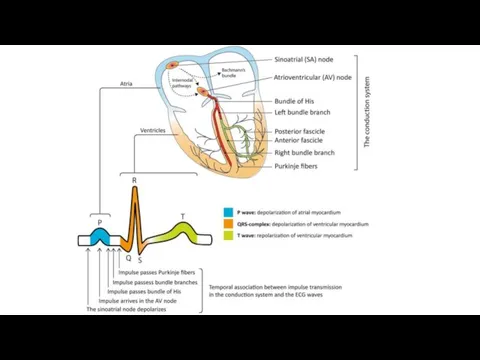
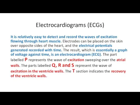
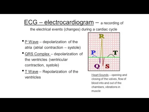
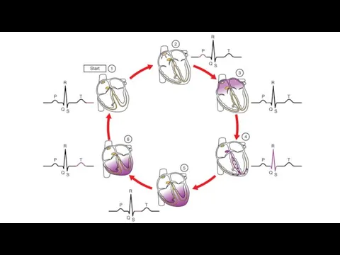
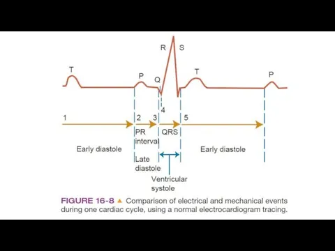
- 15. Electrocardiograms (ECGs) It is relatively easy to detect and record the waves of excitation flowing through
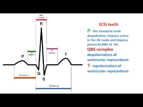
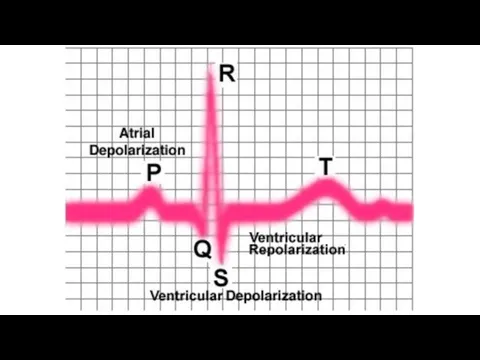
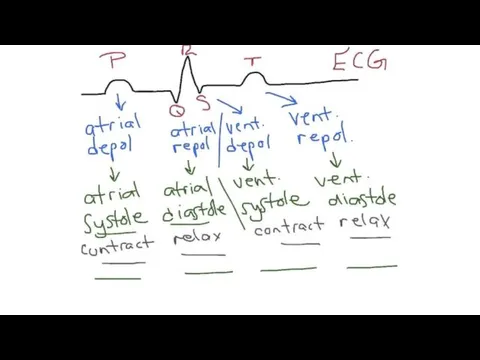
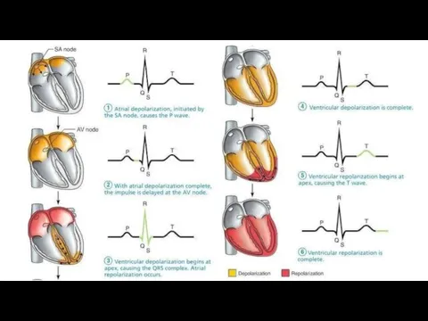
- 17. ECG teeth P- the sinoatrial node despoliation, impulse arrive in the AV node and impulse passes
- 27. Скачать презентацию
The cardiac cycle and ECG
The cardiac cycle and ECG
Learning objectives
11.1.3.4 use an electrocardiogram to describe the cardiac cycle
11.1.3.5
Learning objectives
11.1.3.4 use an electrocardiogram to describe the cardiac cycle
11.1.3.5
Success creteria
1.Investigate the electrical process of in the heart .
Success creteria
1.Investigate the electrical process of in the heart .
Terminology
Atrium, ventricle, contract, systole, blood pressure, valves, aorta, pulmonary vein
Terminology
Atrium, ventricle, contract, systole, blood pressure, valves, aorta, pulmonary vein

Cardiac cycle
Your heart beats around 70 times a minute. The cardiac
Cardiac cycle
Your heart beats around 70 times a minute. The cardiac
Atrial systole
The heart is filled with blood and the muscle
Atrial systole
The heart is filled with blood and the muscle
Ventricular systole
About 0.1 seconds after the atria contract, the ventricles contract.
Ventricular systole
About 0.1 seconds after the atria contract, the ventricles contract.
Ventricular diastole
Ventricular systole lasts for about 0.3 seconds. The muscle
Ventricular diastole
Ventricular systole lasts for about 0.3 seconds. The muscle
Deoxygenated blood from body to RA through vena cava
Blood from RA
Deoxygenated blood from body to RA through vena cava
Blood from RA
Electrocardiograms (ECGs)
It is relatively easy to detect and record the waves
Electrocardiograms (ECGs)
It is relatively easy to detect and record the waves
ECG teeth
P- the sinoatrial node despoliation, impulse arrive in the AV
ECG teeth
P- the sinoatrial node despoliation, impulse arrive in the AV

 Биомеханика и механика. Биологические основы ортодонтического лечения
 Условные предложения. Виды условных предложений
 Трансфузиология. Гемодиализ
 Зәр шығару және сыртқы жыныс жүйесінің физикалық тексеру әдістері. Балалардағы ерекшелігі
 Ответ острой фазы. Лихорадка
 Современные методы лечения скрытого немелкоклеточного рака легкого (НМРЛ)
 Эпидемиология, клиника, профилактика ВИЧ-инфекции
 Внелегочный туберкулез. Туберкулезный менингит
 Медицинская консультация 2010. 2 часть
 Рекомендации по диагностике и лечению хронического панкреатита
 Инновационные методы диагностики ревматических заболеваний
 Остеохондропатии и остеодистрофии
 Современные тенденции в интенсивной терапии травматического кровотечения
 Острый панкреатит
 Особенности течения и лечение бронхиальной астмы у беременных
 Why do we care about goat reproduction
 Отчет о деятельности ГБУ Каргапольская ЦРБ
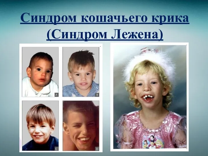 Синдром кошачьего крика (Синдром Лежена)
 Нарушение водно-солевого обмена. Тема 6
 Стоматологиялық науқастарды функционалдық тексеру әдістері
 Рак печени
 Гигиенические требования к освещению детских и подростковых организаций, к санитарно-техническому оборудованию
 Заболевания дыхательной системы: виды и особенности
 Мы против наркотиков
 Лайм-боррелиоз
 Цитокины. Основные компоненты системы цитокинов
 Общая и частная психопатология
 Pharmaceutical monitoring and evaluation