- Radiation diagnosis of acute and chronic heart failure

Содержание
- 2. Heart failure • Acute heart failure - develops very quickly (from several minutes to several hours).
- 3. • Chronic heart failure - the formation of pathology is gradual and develops throughout the weeks,
- 4. There are three types of lesion localization: 1. Left ventricular heart failure 2. Right ventricular heart
- 5. X-ray examination. In acute or uncompensated chronic heart failure, chest radiographic examination of the chest may
- 6. Chest X-ray can help in diagnosing augmentation of the left atrium with mitral valve defects, calcification
- 7. An increase in the right heart with pulmonary hypertension. Direct projection. Signs of an increase in
- 8. "Trapezoid" configuration of the shadow of the heart: cardiomegaly in severe heart failure. The shadow of
- 9. Radiograph of the heart with an increase in the left ventricle. Aortic configuration of the heart.
- 10. Radiography of the heart with an increase in the left atrium. Mitral configuration of the heart.
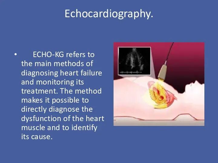
- 11. Echocardiography. ECHO-KG refers to the main methods of diagnosing heart failure and monitoring its treatment. The
- 12. Doppler echocardiography allows to identify and assess valve stenosis and regurgitation, congenital heart defects, valvular vegetations,
- 13. Transthoracic echocardiography. Defect of interventricular septum. Duplex study. There is a hole in the interventricular septum
- 15. Скачать презентацию
Heart failure
• Acute heart failure
- develops very quickly (from several minutes
Heart failure
• Acute heart failure
- develops very quickly (from several minutes
• Chronic heart failure
- the formation of pathology is gradual and
• Chronic heart failure
- the formation of pathology is gradual and
There are three types of lesion localization:
1. Left ventricular heart failure
2.
There are three types of lesion localization:
1. Left ventricular heart failure
2.
X-ray examination.
In acute or uncompensated chronic heart failure, chest radiographic examination
X-ray examination.
In acute or uncompensated chronic heart failure, chest radiographic examination
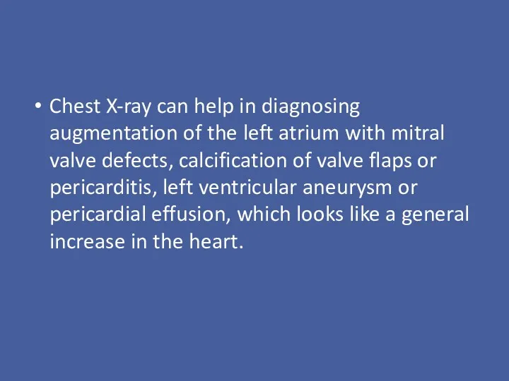
Chest X-ray can help in diagnosing augmentation of the left atrium
Chest X-ray can help in diagnosing augmentation of the left atrium
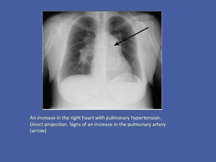
An increase in the right heart with pulmonary hypertension. Direct projection.
An increase in the right heart with pulmonary hypertension. Direct projection.
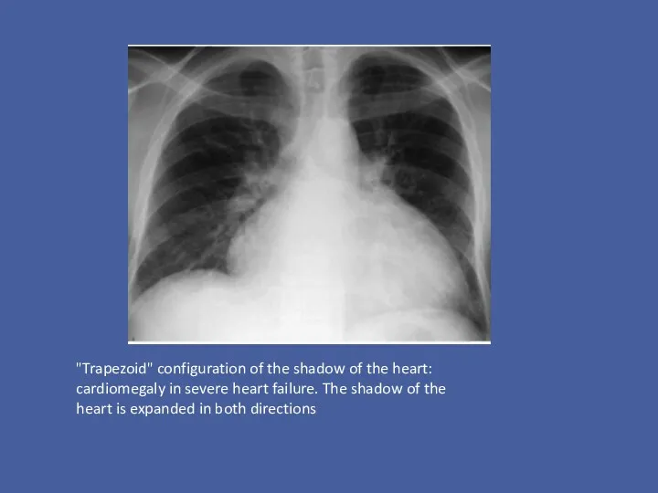
"Trapezoid" configuration of the shadow of the heart: cardiomegaly in severe
"Trapezoid" configuration of the shadow of the heart: cardiomegaly in severe
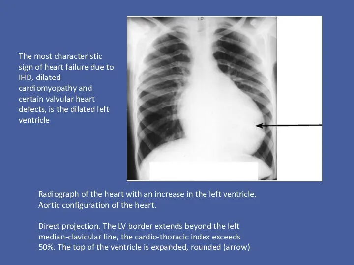
Radiograph of the heart with an increase in the left ventricle.
Radiograph of the heart with an increase in the left ventricle.
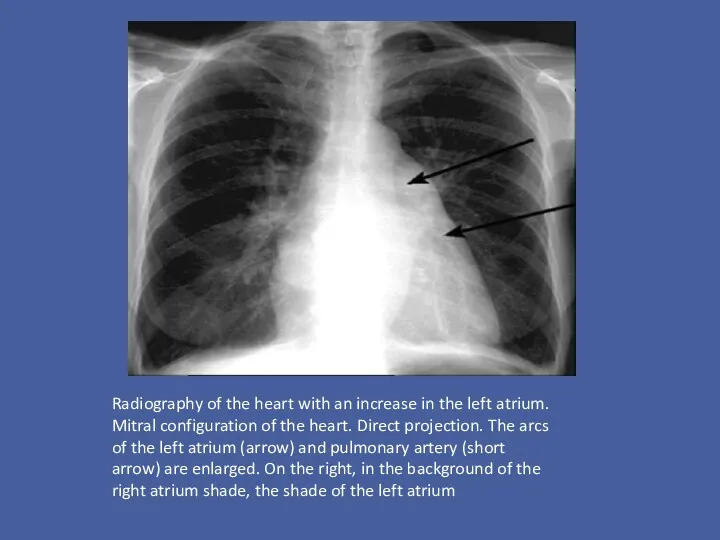
Radiography of the heart with an increase in the left atrium.
Radiography of the heart with an increase in the left atrium.
Echocardiography.
ECHO-KG refers to the main methods of diagnosing heart failure
Echocardiography.
ECHO-KG refers to the main methods of diagnosing heart failure
Doppler echocardiography allows to identify and assess valve stenosis and regurgitation,
Doppler echocardiography allows to identify and assess valve stenosis and regurgitation,
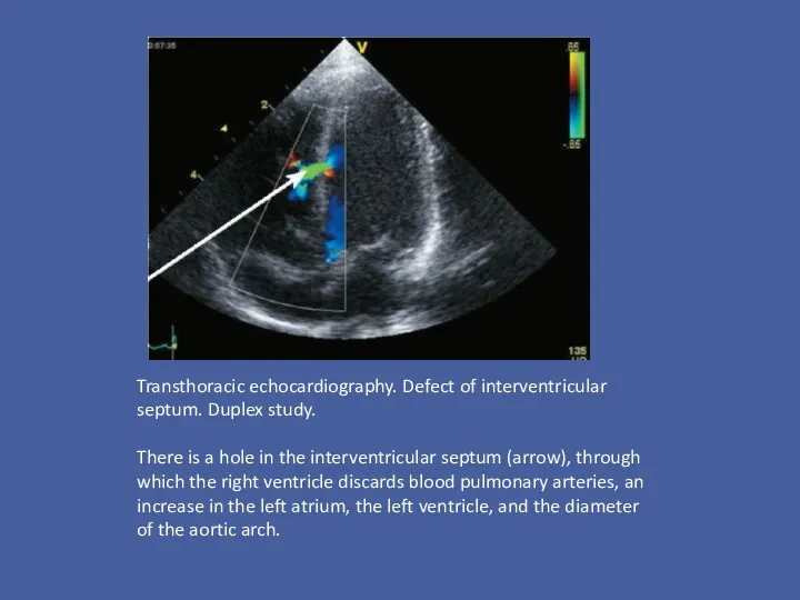
Transthoracic echocardiography. Defect of interventricular septum. Duplex study.
There is a hole
Transthoracic echocardiography. Defect of interventricular septum. Duplex study.
There is a hole
 Нарушения речи и особенности их коррекции у детей с интеллектуальной недостаточностью
 Лікарські засоби, що впливають на серцево-судинну систему та функцію нирок
 Геморрагические лихорадки
 Гемолитическая болезнь плода и новорожденного. Повышение непрямого билирубина
 Особенности фармакокинетики лекарственных веществ, вводимых роженице в родах и их влияние на плод
 Инвазия. Формы инвазии
 Расспрос больных с заболеваниями органов дыхания. Общий осмотр. Осмотр и пальпация грудной клетки. Перкуссия легких
 Әлеуметтік диагностика
 Энергетическая ценность пищевых продуктов
 Функционирование учреждения здравоохранения в условиях чрезвычайных ситуаций
 Клиническая психология
 Предупреждение желудочно-кишечных болезней
 Пролиферативное воспаление
 Переломи і опіки
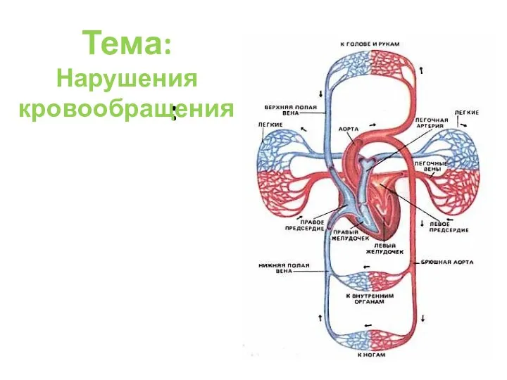 ОП нарушения кровообращения
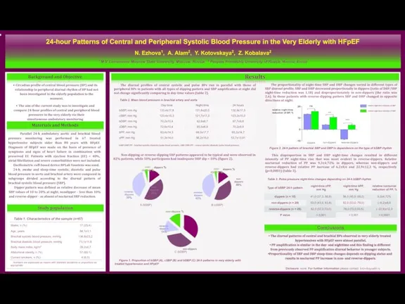 24-hour patterns of central and peripheral systolic blood pressure in the very elderly with HFpEF
 Периферическая нервная система. Спинномозговые нервы
 Медицинская служба Вооруженных сил РФ в ЧС
 Рожистое воспаление
 Гипогликемия и гипергликемия у новорожденных
 Первая помощь при термотравмах и электротравмах
 Переливание крови. История. Изогемагглютинация, группы крови. Механизм действия перелитой крови
 Проблема инвалидности
 Эффект применения Флуфеназина в лечении синдрома Туретта
 Синдром пангипопитуитаризма
 Вторичный метаболизм
 Клинико-психологическое сопровождение пожилых людей страдающих инволлюционным паранойдом
 Ультразвуковые маркеры хромосомной патологии