- Warm-Up

Содержание
- 2. Chapter 42 – Part I Circulation
- 3. What you need to know: Circulatory vessels, heart chambers, route of mammalian circulation Evolution of the
- 4. Transport systems (circulation) linked with gas exchange (respiration) Diffusion of gases only rapid across small distances
- 5. Circulatory System = Blood + Vessels + Heart Open circulatory system: blood bathes organs directly Blood
- 6. Figure 42.10a Endothelium Artery Smooth muscle Connective tissue Capillary Valve Vein Basal lamina Endothelium Smooth muscle
- 7. Types of Blood Vessels arterioles venules
- 8. Blood enters through an atrium and is pumped out through a ventricle Fish = single circulation
- 9. Double circulation pathways in vertebrates
- 10. Pathway of blood through heart
- 11. Superior vena cava Pulmonary artery Capillaries of right lung Pulmonary vein Aorta Inferior vena cava Right
- 12. Cardiac cycle Systole: contraction or pumping phase Diastole: relaxation or filling phase Heart rate: # beats/minute
- 13. Figure 42.8-3 0.1 sec 0.4 sec 0.3 sec 2 1 3
- 14. Valves: prevent backflow of blood The atrioventricular (AV) valves (tricuspid, bicuspid) separate each atrium and ventricle
- 15. Sinoatrial (SA) node: pacemaker of heart, in right atrium
- 16. The pacemaker is regulated by two portions of the nervous system: the sympathetic and parasympathetic divisions
- 17. Blood Pressure BP = systolic/diastolic pressure Systolic: heart contracts Diastolic: heart relaxed Normal: 120/70 Pulse: rhythmic
- 18. Blood pressure reading: 120/70 120 70 Sounds stop Sounds audible in stethoscope 120 Artery closed 1
- 19. Direction of blood flow in vein (toward heart) Valve (open) Skeletal muscle Valve (closed) Figure 42.13
- 20. Lymphatic System: returns lost fluid and proteins to blood as lymph Lymph Nodes: filter lymph, house
- 21. Blood Plasma (55%) – water, ions, proteins, gases, nutrients, wastes, hormones Cells (45%) – RBC, WBC,
- 22. Figure 42.17 Plasma 55% Constituent Major functions Water Ions (blood electrolytes) Sodium Potassium Calcium Magnesium Chloride
- 23. Figure 42.18 Collagen fibers 1 2 3 Platelet Platelet plug Fibrin clot Fibrin clot formation Red
- 24. Cardiovascular Disease Atherosclerosis: buildup of plaque deposits within arteries Heart attack (myocardial infarction): blockage of one
- 26. Скачать презентацию
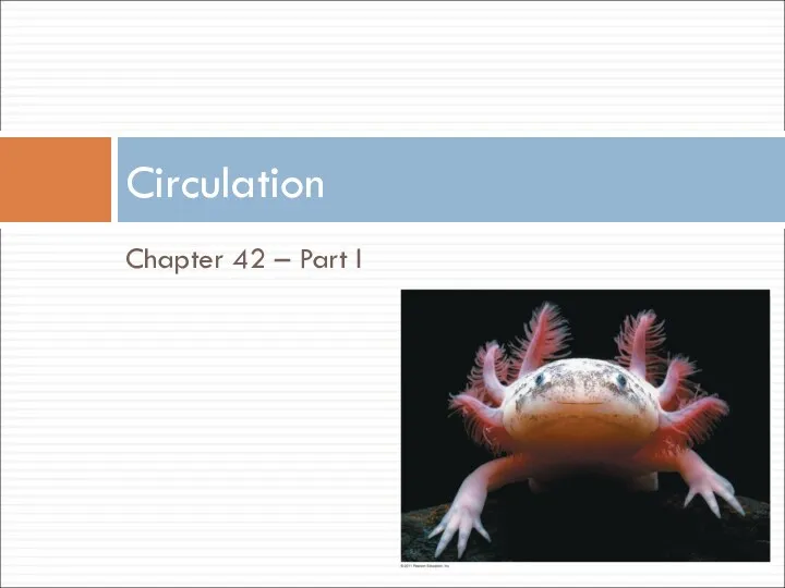
Chapter 42 – Part I
Circulation
Chapter 42 – Part I
Circulation
What you need to know:
Circulatory vessels, heart chambers, route of mammalian
What you need to know:
Circulatory vessels, heart chambers, route of mammalian
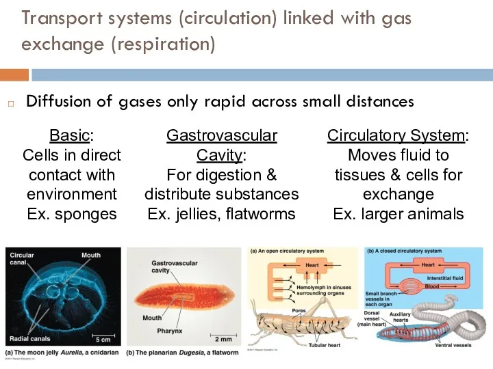
Transport systems (circulation) linked with gas exchange (respiration)
Diffusion of gases only
Transport systems (circulation) linked with gas exchange (respiration)
Diffusion of gases only
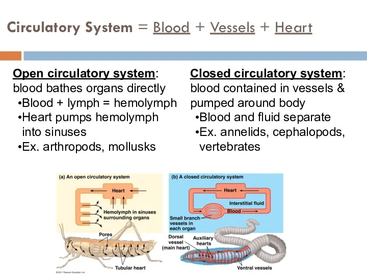
Circulatory System = Blood + Vessels + Heart
Open circulatory system: blood
Circulatory System = Blood + Vessels + Heart
Open circulatory system: blood
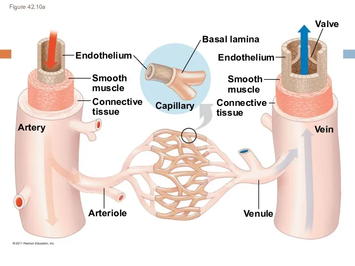
Figure 42.10a
Endothelium
Artery
Smooth
muscle
Connective
tissue
Capillary
Valve
Vein
Basal lamina
Endothelium
Smooth
muscle
Connective
tissue
Venule
Arteriole
Figure 42.10a
Endothelium
Artery
Smooth
muscle
Connective
tissue
Capillary
Valve
Vein
Basal lamina
Endothelium
Smooth
muscle
Connective
tissue
Venule
Arteriole
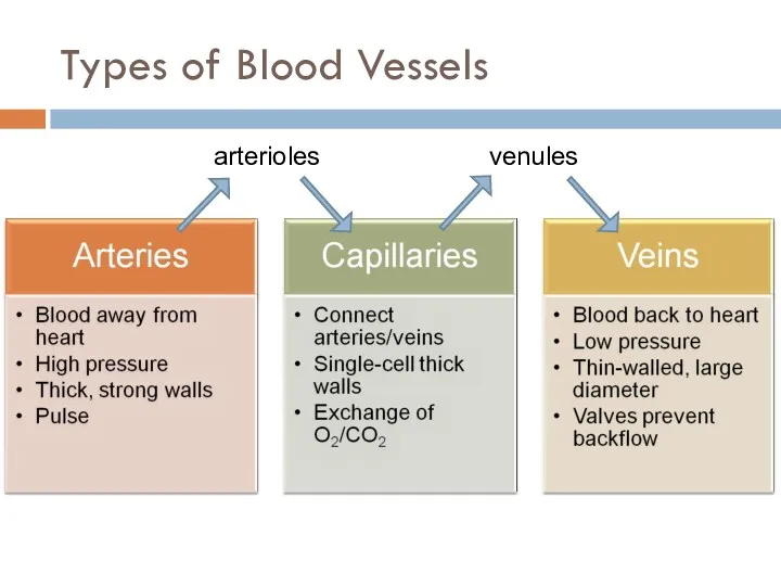
Types of Blood Vessels
arterioles
venules
Types of Blood Vessels
arterioles
venules
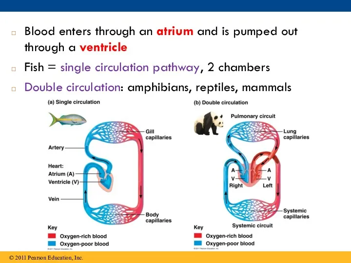
Blood enters through an atrium and is pumped out through a
Blood enters through an atrium and is pumped out through a
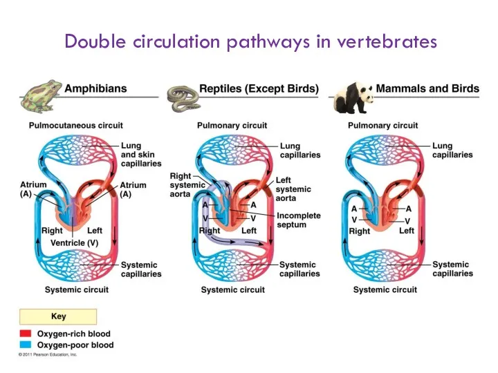
Double circulation pathways in vertebrates
Double circulation pathways in vertebrates
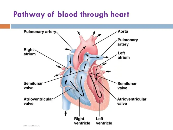
Pathway of blood through heart
Pathway of blood through heart
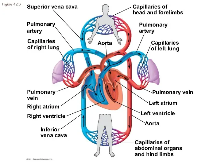
Superior vena cava
Pulmonary
artery
Capillaries
of right lung
Pulmonary
vein
Aorta
Inferior
vena cava
Right ventricle
Capillaries of
abdominal organs
and hind limbs
Right
Superior vena cava
Pulmonary
artery
Capillaries
of right lung
Pulmonary
vein
Aorta
Inferior
vena cava
Right ventricle
Capillaries of
abdominal organs
and hind limbs
Right
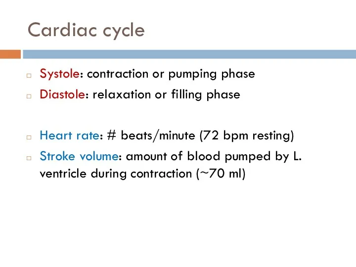
Cardiac cycle
Systole: contraction or pumping phase
Diastole: relaxation or filling phase
Heart rate:
Cardiac cycle
Systole: contraction or pumping phase
Diastole: relaxation or filling phase
Heart rate:
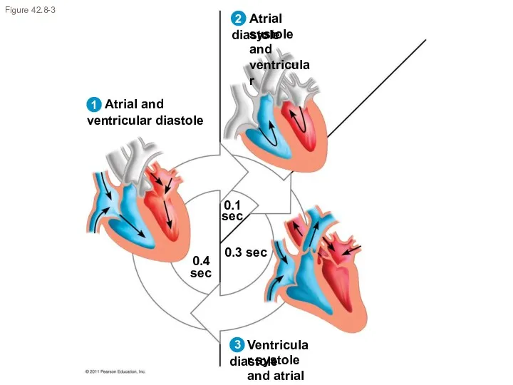
Figure 42.8-3
0.1
sec
0.4
sec
0.3 sec
2
1
3
Figure 42.8-3
0.1
sec
0.4
sec
0.3 sec
2
1
3
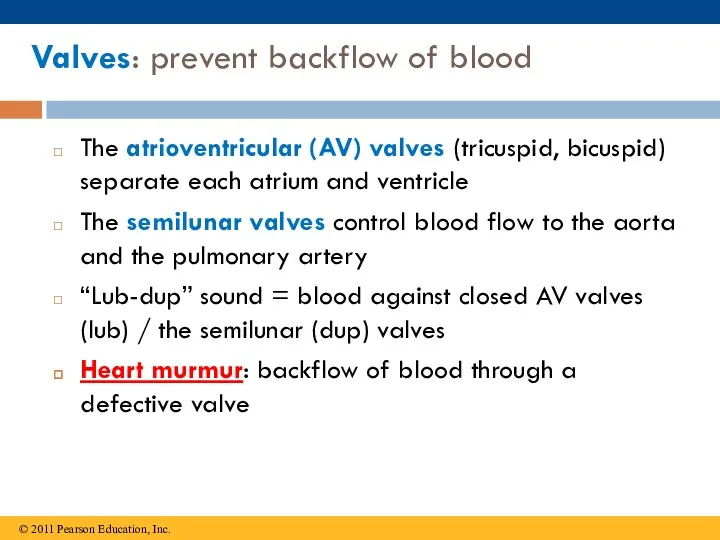
Valves: prevent backflow of blood
The atrioventricular (AV) valves (tricuspid, bicuspid) separate
Valves: prevent backflow of blood
The atrioventricular (AV) valves (tricuspid, bicuspid) separate
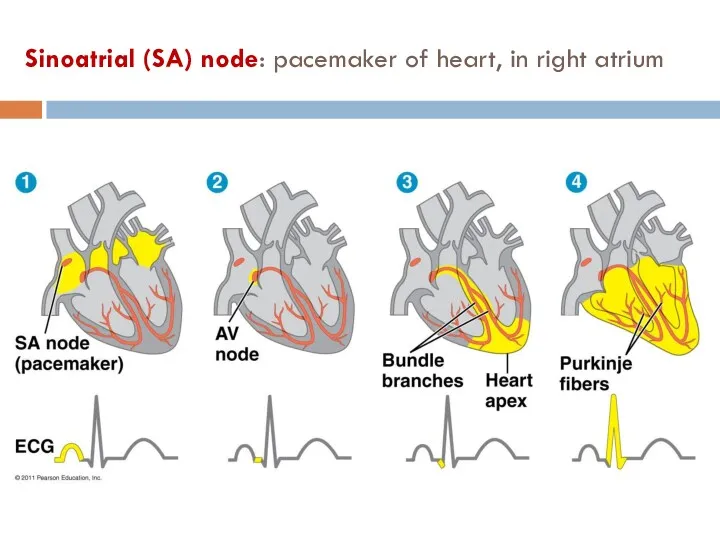
Sinoatrial (SA) node: pacemaker of heart, in right atrium
Sinoatrial (SA) node: pacemaker of heart, in right atrium
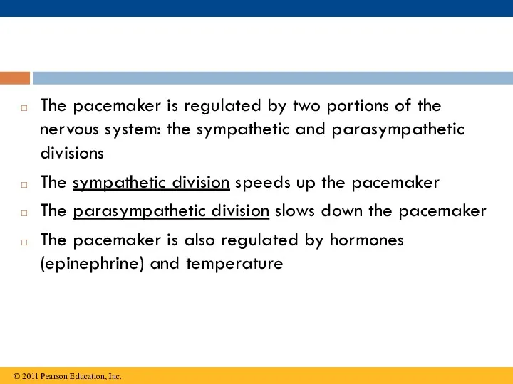
The pacemaker is regulated by two portions of the nervous system:
The pacemaker is regulated by two portions of the nervous system:
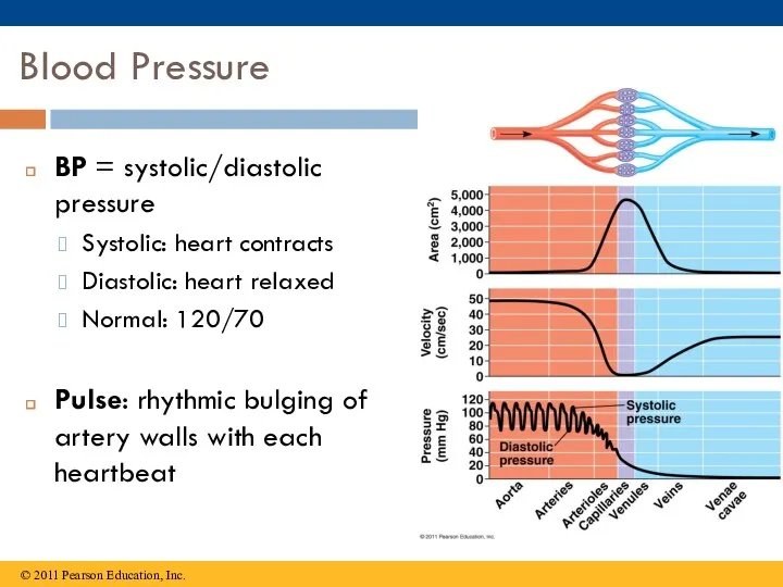
Blood Pressure
BP = systolic/diastolic pressure
Systolic: heart contracts
Diastolic: heart relaxed
Normal: 120/70
Pulse: rhythmic
Blood Pressure
BP = systolic/diastolic pressure
Systolic: heart contracts
Diastolic: heart relaxed
Normal: 120/70
Pulse: rhythmic
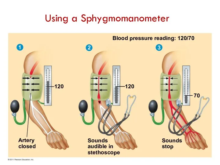
Blood pressure reading: 120/70
120
70
Sounds
stop
Sounds
audible in
stethoscope
120
Artery
closed
1
2
3
Using a Sphygmomanometer
Blood pressure reading: 120/70
120
70
Sounds
stop
Sounds
audible in
stethoscope
120
Artery
closed
1
2
3
Using a Sphygmomanometer
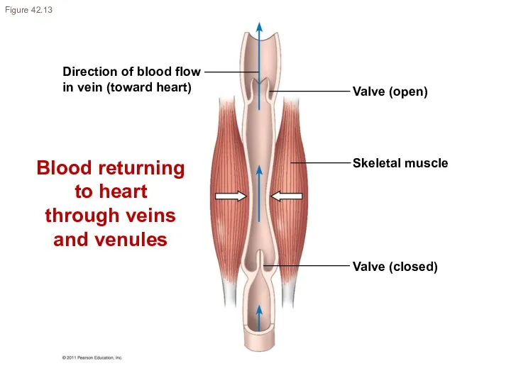
Direction of blood flow
in vein (toward heart)
Valve (open)
Skeletal muscle
Valve (closed)
Figure 42.13
Blood
Direction of blood flow
in vein (toward heart)
Valve (open)
Skeletal muscle
Valve (closed)
Figure 42.13
Blood
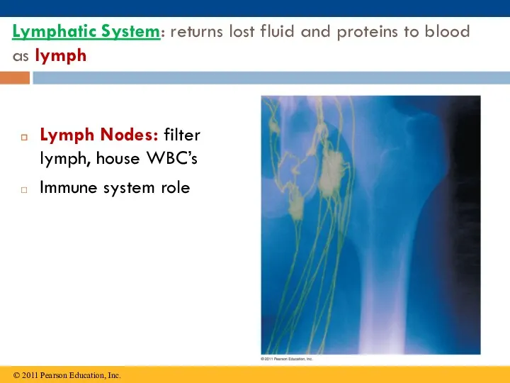
Lymphatic System: returns lost fluid and proteins to blood as lymph
Lymph
Lymphatic System: returns lost fluid and proteins to blood as lymph
Lymph
Blood
Plasma (55%) – water, ions, proteins, gases, nutrients, wastes, hormones
Cells (45%)
Blood
Plasma (55%) – water, ions, proteins, gases, nutrients, wastes, hormones
Cells (45%)
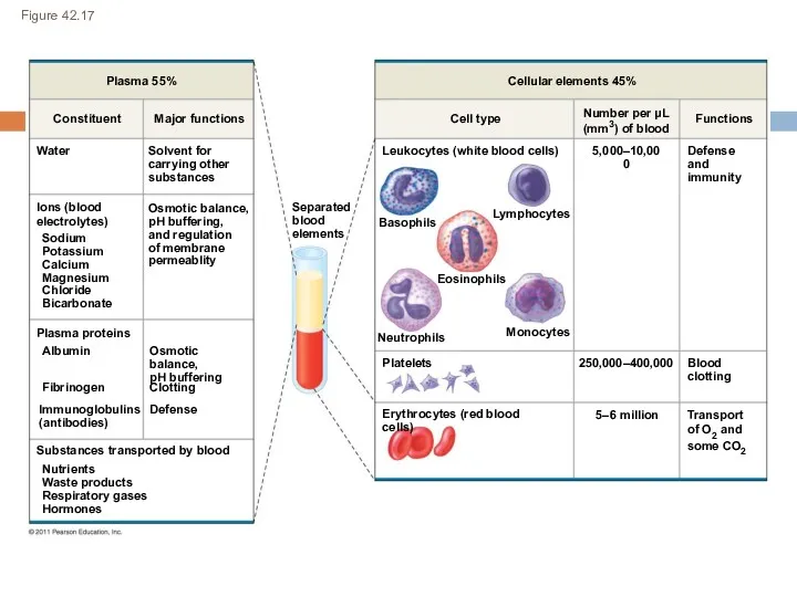
Figure 42.17
Plasma 55%
Constituent
Major functions
Water
Ions (blood
electrolytes)
Sodium
Potassium
Calcium
Magnesium
Chloride
Bicarbonate
Solvent for
carrying other
substances
Osmotic balance,
pH buffering,
and regulation
of membrane
permeablity
Plasma
Figure 42.17
Plasma 55%
Constituent
Major functions
Water
Ions (blood
electrolytes)
Sodium
Potassium
Calcium
Magnesium
Chloride
Bicarbonate
Solvent for
carrying other
substances
Osmotic balance,
pH buffering,
and regulation
of membrane
permeablity
Plasma
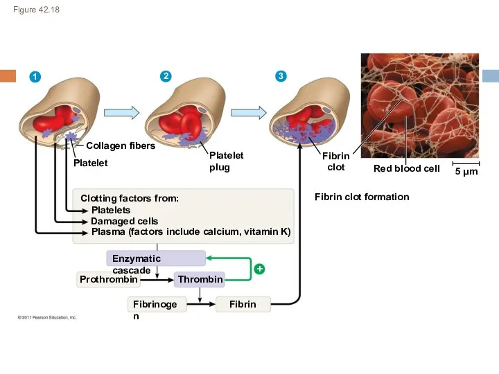
Figure 42.18
Collagen fibers
1
2
3
Platelet
Platelet
plug
Fibrin
clot
Fibrin clot formation
Red blood cell
5 μm
Clotting factors from:
Platelets
Damaged cells
Plasma
Figure 42.18
Collagen fibers
1
2
3
Platelet
Platelet
plug
Fibrin
clot
Fibrin clot formation
Red blood cell
5 μm
Clotting factors from:
Platelets
Damaged cells
Plasma
Cardiovascular Disease
Atherosclerosis: buildup of plaque deposits within arteries
Heart attack (myocardial infarction):
Cardiovascular Disease
Atherosclerosis: buildup of plaque deposits within arteries
Heart attack (myocardial infarction):
 Биогенді (s.p.d) элементтермен олардың қосылыстарының медициналық және биологиялық маңызы
 Кислоты, их состав и названия
 Карбоновые кислоты и их функциональные производные
 Ароматические углеводороды
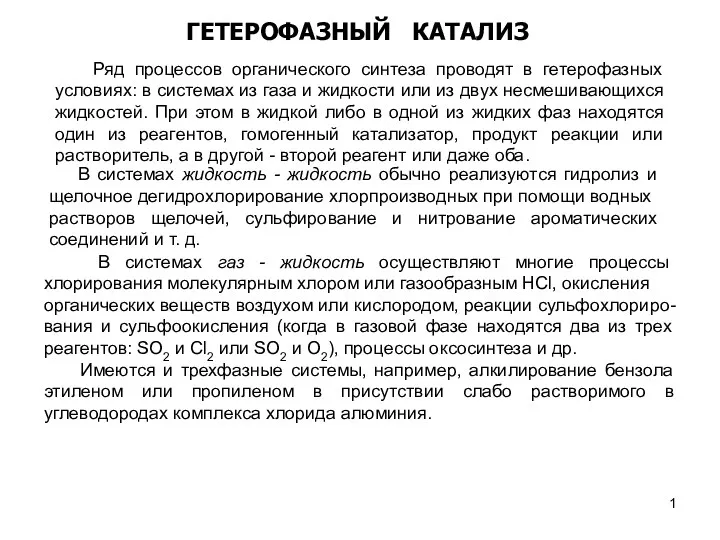 Гетерофазный катализ. (Лекция 20)
 Горение топлива
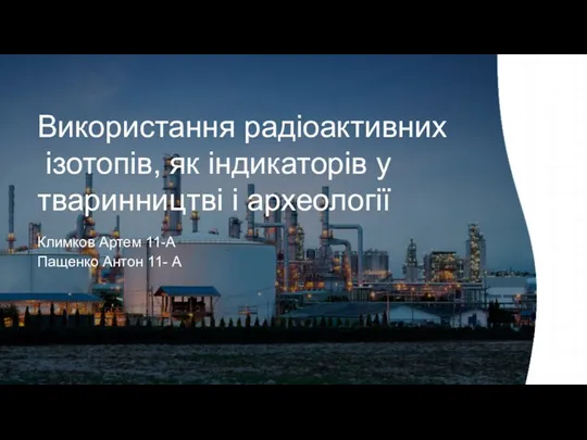 Використання радіоактивних ізотопів, як індикаторів у тваринництві і археології
 Основные классы неорганических соединений
 Соли, их классификация и свойства
 Органічна хімія
 Розв’язування задач за рівняннями реакцій з використанням розчинів із певною масовою часткою розчиненої речовини. Урок 13-14
 Жёсткость воды
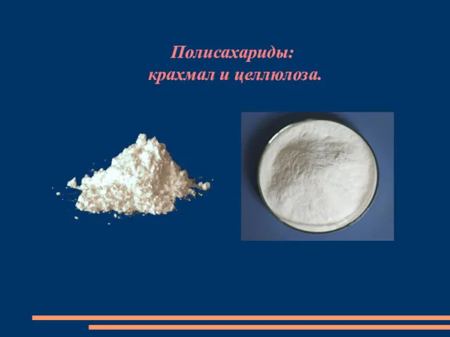 Полисахариды: крахмал и целлюлоза
 Растворы. Часть 2. Лекция №7
 Химия атмосферы. Химические процессы в тропосфере
 Буферные системы
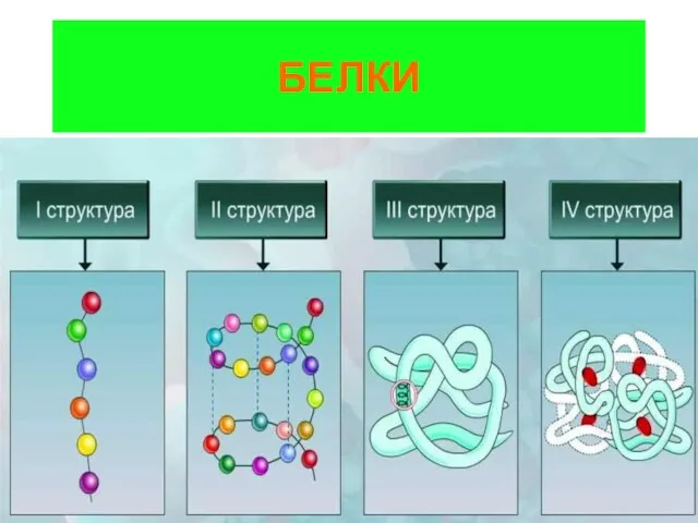 Белки. Строение
 Классификация химических элементов. Составитель. 8 класс
 Минералы для ИЗБ
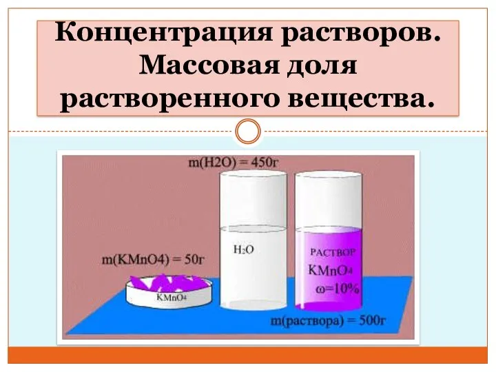 Концентрация растворов. Массовая доля растворенного вещества. Урок 1
 Химический элемент медь
 Кинетика химических реакций
 Кремний и его соединения
 Галогены. Расположите галогены в порядке их открытия
 Важнейшие реакции в органической химии
 Технология производства аминоальдегидных смол
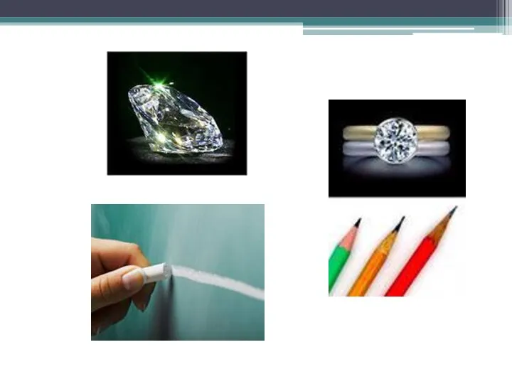 Углерод. Физические и химические свойства
 Гидролиз солей